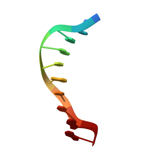
Structure of the B-DNA decamer C-C-A-A-C-I-T-T-G-G in two different space groups: conformational flexibility of B-DNA.
Lipanov, A., Kopka, M.L., Kaczor-Grzeskowiak, M., Quintana, J., Dickerson, R.E.(1993) Biochemistry 32: 1373-1389
- PubMed: 8448146 Search on PubMed
- DOI: https://doi.org/10.1021/bi00056a024
- Primary Citation Related Structures:
1D60, 1D61, 1D62 - PubMed Abstract:
For the first time, the same B-DNA oligomer has been crystallized and its structure solved in two different space groups. Crystallization of C-C-A-A-C-I-T-T-G-G with Ca2+ yields monoclinic space group C2 with a = 31.87 A, b = 25.69 A, c = 34.21 A, beta = 114.1 degrees, and five base pairs per asymmetric unit. The 5026 2 sigma data to 1.3 A refine to R = 0.152 with 72 waters, one heptavalent hydrated calcium complex, and one cacodylate ion per asymmetric unit. In contrast, crystallization with Mg2+ yields trigonal space group P3(2)21 with a = b = 33.23 A, c = 94.77 A, gamma = 120 degrees, and 10 base pairs per asymmetric unit. The 1725 2 sigma data to 2.2 A refine to R = 0.164 with 36 water molecules and one octahedral magnesium complex per asymmetric unit. The monoclinic form is virtually isostructural with previously solved monoclinic decamers, including twist angles of ca. 50 degrees at C-A and T-G steps. In contrast, the trigonal structure has quite different local helix parameters, with twist angles of ca. 36 degrees at the corresponding steps. These local parameter differences can only be attributed to crystal packing, suggesting that certain sequences of B-DNA are more flexible and influenced by their surroundings than had previously been thought. Such deformability may be important for interaction of B-DNA with control proteins, where both static structure and dynamic deformability comprise components of the recognition process. The crossing of two helices at an angle of 120 degrees in the trigonal cell is a model for an antiparallel, uncrossed Holliday junction, as has been noted earlier by Timsit and Moras [Timsit, Y., & Moras, D. (1991) J. Mol. Biol. 221, 919-940] from a rhombohedral DNA dodecamer structure analysis.
- Department of Chemistry and Biochemistry, University of California, Los Angeles 90024.
Organizational Affiliation: