1D2F | pdb_00001d2f

X-RAY STRUCTURE OF MALY FROM ESCHERICHIA COLI: A PYRIDOXAL-5'-PHOSPHATE-DEPENDENT ENZYME ACTING AS A MODULATOR IN MAL GENE EXPRESSION


Experimental Data Snapshot

  • Method: X-RAY DIFFRACTION
  • Resolution: 2.50 Å
  • R-Value Free: 
    0.262 (Depositor), 0.252 (DCC) 
  • R-Value Work: 
    0.201 (Depositor), 0.198 (DCC) 
  • R-Value Observed: 
    0.204 (Depositor) 

wwPDB Validation 3D Report Full Report

Validation slider image for 1D2F

Ligand Structure Quality Assessment 


This is version 1.3 of the entry. See complete history

Re-refinement Note

A newer entry is available that reflects an alternative modeling of the original data: 8BOB

Literature

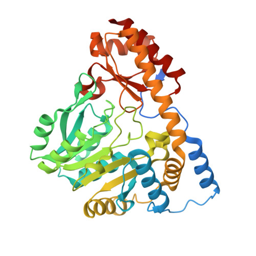
X-ray structure of MalY from Escherichia coli: a pyridoxal 5'-phosphate-dependent enzyme acting as a modulator in mal gene expression.

Clausen, T.Schlegel, A.Peist, R.Schneider, E.Steegborn, C.Chang, Y.S.Haase, A.Bourenkov, G.P.Bartunik, H.D.Boos, W.

(2000) EMBO J 19: 831-842

  • DOI: https://doi.org/10.1093/emboj/19.5.831
  • Primary Citation Related Structures: 
    1D2F

  • PubMed Abstract: 

    MalY represents a bifunctional pyridoxal 5'-phosphate-dependent enzyme acting as a beta-cystathionase and as a repressor of the maltose regulon. Here we present the crystal structures of wild-type and A221V mutant protein. Each subunit of the MalY dimer is composed of a large pyridoxal 5'-phosphate-binding domain and a small domain similar to aminotransferases. The structural alignment with related enzymes identifies residues that are generally responsible for beta-lyase activity and depicts a unique binding mode of the pyridoxal 5'-phosphate correlated with a larger, more flexible substrate-binding pocket. In a screen for MalY mutants with reduced mal repressor properties, mutations occurred in three clusters: I, 83-84; II, 181-189 and III, 215-221, which constitute a clearly distinguished region in the MalY crystal structure far away from the cofactor. The tertiary structure of one of these mutants (A221V) demonstrates that positional rearrangements are indeed restricted to regions I, II and III. Therefore, we propose that a direct protein-protein interaction with MalT, the central transcriptional activator of the maltose system, underlies MalY-dependent repression of the maltose system.


  • Organizational Affiliation
    • Max-Planck-Institut für Biochemie, Abteilung Strukturforschung, D-82152 Martinsried.

Macromolecule Content 

  • Total Structure Weight: 87.86 kDa 
  • Atom Count: 5,914 
  • Modeled Residue Count: 729 
  • Deposited Residue Count: 780 
  • Unique protein chains: 1

Macromolecules

Find similar proteins by:|  3D Structure
Entity ID: 1
MoleculeChains  Sequence LengthOrganismDetailsImage
MALY PROTEIN
A, B
390Escherichia coliMutation(s): 0 
EC: 4.4.1.13
UniProt
Find proteins for P23256 (Escherichia coli (strain K12))
Explore P23256 
Go to UniProtKB:  P23256
Entity Groups
Sequence Clusters30% Identity50% Identity70% Identity90% Identity95% Identity100% Identity
UniProt GroupP23256
Sequence Annotations
Expand
Reference Sequence

Experimental Data & Validation

Experimental Data

  • Method: X-RAY DIFFRACTION
  • Resolution: 2.50 Å
  • R-Value Free:  0.262 (Depositor), 0.252 (DCC) 
  • R-Value Work:  0.201 (Depositor), 0.198 (DCC) 
  • R-Value Observed: 0.204 (Depositor) 
Space Group: C 2 2 21
Unit Cell:
Length ( Å )Angle ( ˚ )
a = 57.51α = 90
b = 107.19β = 90
c = 256.4γ = 90
Software Package:
Software NamePurpose
MLPHAREphasing
X-PLORrefinement
MOSFLMdata reduction
CCP4data scaling

Structure Validation

View Full Validation Report


Ligand Structure Quality Assessment 


Entry History 

Deposition Data

  • Released Date: 2000-01-12 
  • Deposition Author(s): Clausen, T.

Revision History  (Full details and data files)

  • Version 1.0: 2000-01-12
    Type: Initial release
  • Version 1.1: 2008-04-27
    Changes: Version format compliance
  • Version 1.2: 2011-07-13
    Changes: Non-polymer description, Version format compliance
  • Version 1.3: 2025-03-26
    Changes: Data collection, Database references, Derived calculations, Structure summary