1CTF

STRUCTURE OF THE C-TERMINAL DOMAIN OF THE RIBOSOMAL PROTEIN L7/L12 FROM ESCHERICHIA COLI AT 1.7 ANGSTROMS


Experimental Data Snapshot

  • Method: X-RAY DIFFRACTION
  • Resolution: 1.70 Å
  • R-Value Work: 0.174 

wwPDB Validation   3D Report Full Report


This is version 1.3 of the entry. See complete history


Literature

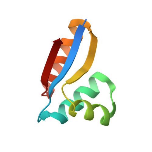
Structure of the C-terminal domain of the ribosomal protein L7/L12 from Escherichia coli at 1.7 A.

Leijonmarck, M.Liljas, A.

(1987) J Mol Biol 195: 555-579

  • DOI: https://doi.org/10.1016/0022-2836(87)90183-5
  • Primary Citation of Related Structures:  
    1CTF

  • PubMed Abstract: 
  • The structure of a C-terminal fragment of the ribosomal protein L7/L12 from Escherichia coli has been refined using crystallographic data to 1.7 A resolution. The R-value is 17.4%. Six residues at the N terminus are too disordered in the structure to be localized ...

    The structure of a C-terminal fragment of the ribosomal protein L7/L12 from Escherichia coli has been refined using crystallographic data to 1.7 A resolution. The R-value is 17.4%. Six residues at the N terminus are too disordered in the structure to be localized. These residues are probably part of a hinge in the complete L7/L12 molecule. The possibility that a 2-fold crystallographic axis is a molecular 2-fold axis is discussed. A patch of invariant residues on the surface of the dimer is probably involved in functional interactions with elongation factors.


    Related Citations: 
    • Structural Studies on the Protein L7(Slash)L12 from E. Coli Ribosomes
      Leijonmarck, M., Pettersson, I., Liljas, A.
      (1981) Structural Aspects Of Recognition And Assembly In Biological Macromolecules --: 761
    • Crystal Structure of a Ribosomal Component at 2.6 Angstroms Resolution
      Leijonmarck, M., Eriksson, S., Liljas, A.
      (1980) Nature 286: 824
    • Isolation and Crystallization of Stable Domains of the Protein L7(Slash)L12 from Escherichia Coli Ribosomes
      Liljas, A., Eriksson, S., Donner, D., Kurland, C.G.
      (1978) FEBS Lett 88: 300

    Organizational Affiliation

    Institute of Molecular Biology, University of Uppsala, Sweden.


Macromolecules
Find similar proteins by:  (by identity cutoff)  |  3D Structure
Entity ID: 1
MoleculeChainsSequence LengthOrganismDetailsImage
RIBOSOMAL PROTEIN L7/L12A74Escherichia coliMutation(s): 0 
UniProt
Find proteins for P0A7K2 (Escherichia coli (strain K12))
Explore P0A7K2 
Go to UniProtKB:  P0A7K2
Entity Groups  
Sequence Clusters30% Identity50% Identity70% Identity90% Identity95% Identity100% Identity
UniProt GroupP0A7K2
Protein Feature View
Expand
  • Reference Sequence
Small Molecules
Ligands 1 Unique
IDChainsName / Formula / InChI Key2D Diagram3D Interactions
SO4
Query on SO4

Download Ideal Coordinates CCD File 
B [auth A]SULFATE ION
O4 S
QAOWNCQODCNURD-UHFFFAOYSA-L
 Ligand Interaction
Experimental Data & Validation

Experimental Data

  • Method: X-RAY DIFFRACTION
  • Resolution: 1.70 Å
  • R-Value Work: 0.174 
  • Space Group: P 43 21 2
Unit Cell:
Length ( Å )Angle ( ˚ )
a = 54.83α = 90
b = 54.83β = 90
c = 42.67γ = 90
Software Package:
Software NamePurpose
CORELSrefinement

Structure Validation

View Full Validation Report


Entry History 

Deposition Data

Revision History  (Full details and data files)

  • Version 1.0: 1987-01-15
    Type: Initial release
  • Version 1.1: 2008-03-03
    Changes: Version format compliance
  • Version 1.2: 2011-07-13
    Changes: Version format compliance
  • Version 1.3: 2017-11-29
    Changes: Derived calculations, Other