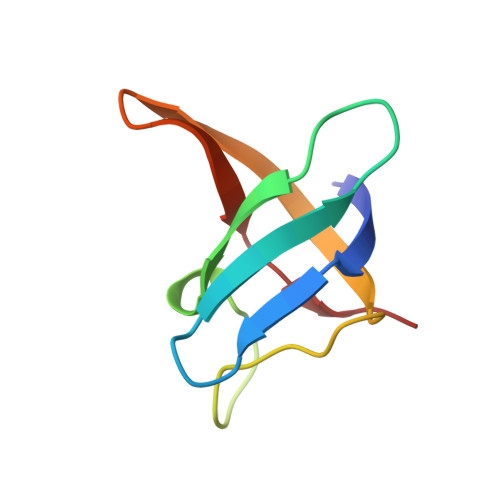
1CSP

CRYSTAL STRUCTURE OF THE BACILLUS SUBTILIS MAJOR COLD SHOCK PROTEIN, CSPB: A UNIVERSAL NUCLEIC-ACID BINDING DOMAIN


Experimental Data Snapshot

  • Method: X-RAY DIFFRACTION
  • Resolution: 2.45 Å
  • R-Value Observed: 0.195 

wwPDB Validation   3D Report Full Report


This is version 1.4 of the entry. See complete history


Literature

Universal nucleic acid-binding domain revealed by crystal structure of the B. subtilis major cold-shock protein.

Schindelin, H.Marahiel, M.A.Heinemann, U.

(1993) Nature 364: 164-168

  • DOI: https://doi.org/10.1038/364164a0
  • Primary Citation of Related Structures:  
    1CSP, 1CSQ

  • PubMed Abstract: 

    The cold-shock response in both Escherichia coli and Bacillus subtilis is induced by an abrupt downshift in growth temperature. It leads to the increased production of the major cold-shock proteins, CS7.4 and CspB, respectively. CS7.4 is a transcriptional activator of two genes. CS7.4 and CspB share 43 per cent sequence identity with the nucleic acid-binding domain of the eukaryotic gene-regulatory Y-box factors. This cold-shock domain is conserved from bacteria to man and contains the RNA-binding RNP1 sequence motif. As a prototype of the cold-shock domain, the structure of CspB has been determined here from two crystal forms. In both, CspB is present as an antiparallel five-stranded beta-barrel. Three consecutive beta-strands, the central one containing the RNP1 motif, create a surface rich in aromatic and basic residues that are presumably involved in nucleic acid binding. Preferential binding of CspB to single-stranded DNA is observed in gel retardation experiments.


  • Organizational Affiliation

    Institut für Kristallographie, Freie Universität Berlin, Germany.


Macromolecules
Find similar proteins by:  (by identity cutoff)  |  3D Structure
Entity ID: 1
MoleculeChains Sequence LengthOrganismDetailsImage
COLD SHOCK PROTEIN B(CSPB)67Bacillus subtilisMutation(s): 0 
UniProt
Find proteins for P32081 (Bacillus subtilis (strain 168))
Explore P32081 
Go to UniProtKB:  P32081
Entity Groups  
Sequence Clusters30% Identity50% Identity70% Identity90% Identity95% Identity100% Identity
UniProt GroupP32081
Sequence Annotations
Expand
  • Reference Sequence
Experimental Data & Validation

Experimental Data

  • Method: X-RAY DIFFRACTION
  • Resolution: 2.45 Å
  • R-Value Observed: 0.195 
  • Space Group: P 32 2 1
Unit Cell:
Length ( Å )Angle ( ˚ )
a = 58.94α = 90
b = 58.94β = 90
c = 46.45γ = 120
Software Package:
Software NamePurpose
TNTrefinement

Structure Validation

View Full Validation Report


Entry History 

Deposition Data

Revision History  (Full details and data files)

  • Version 1.0: 1995-05-12
    Type: Initial release
  • Version 1.1: 2008-03-24
    Changes: Version format compliance
  • Version 1.2: 2011-07-13
    Changes: Version format compliance
  • Version 1.3: 2017-11-29
    Changes: Derived calculations, Other
  • Version 1.4: 2024-02-07
    Changes: Data collection, Database references, Derived calculations