1COW | pdb_00001cow

BOVINE MITOCHONDRIAL F1-ATPASE COMPLEXED WITH AUROVERTIN B


Experimental Data Snapshot

  • Method: X-RAY DIFFRACTION
  • Resolution: 3.10 Å
  • R-Value Free: 
    0.280 (Depositor) 
  • R-Value Work: 
    0.214 (DCC) 

wwPDB Validation 3D Report Full Report

Validation slider image for 1COW

Ligand Structure Quality Assessment 


This is version 1.4 of the entry. See complete history

Literature

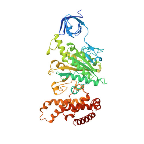
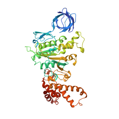
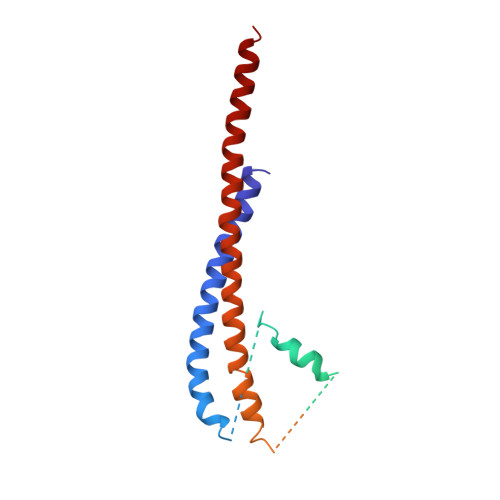
The structure of bovine F1-ATPase complexed with the antibiotic inhibitor aurovertin B.

van Raaij, M.J.Abrahams, J.P.Leslie, A.G.Walker, J.E.

(1996) Proc Natl Acad Sci U S A 93: 6913-6917

  • DOI: https://doi.org/10.1073/pnas.93.14.6913
  • Primary Citation Related Structures: 
    1COW

  • PubMed Abstract: 

    In the structure of bovine mitochondrial F1-ATPase that was previously determined with crystals grown in the presence of adenylyl-imidodiphosphate (AMP-PNP) and ADP, the three catalytic beta-subunits have different conformations and nucleotide occupancies. Adenylyl-imidodiphosphate is bound to one beta-subunit (betaTP), ADP is bound to the second (betaDP), and no nucleotide is bound to the third (betaE). Here we show that the uncompetitive inhibitor aurovertin B binds to bovine F1 at two equivalent sites in betaTP and betaE, in a cleft between the nucleotide binding and C-terminal domains. In betaDP, the aurovertin B pocket is incomplete and is inaccessible to the inhibitor. The aurovertin B bound to betaTP interacts with alpha-Glu399 in the adjacent alphaTP subunit, whereas the aurovertin B bound to betaE is too distant from alphaE to make an equivalent interaction. Both sites encompass betaArg-412, which was shown by mutational studies to be involved in binding aurovertin. Except for minor changes around the aurovertin pockets, the structure of bovine F1-ATPase is the same as determined previously. Aurovertin B appears to act by preventing closure of the catalytic interfaces, which is essential for a catalytic mechanism involving cyclic interconversion of catalytic sites.


  • Organizational Affiliation
    • Medical Research Council Laboratory of Molecular Biology, Cambridge, United Kingdom.

Macromolecule Content 

  • Total Structure Weight: 354.86 kDa 
  • Atom Count: 23,462 
  • Modeled Residue Count: 2,987 
  • Deposited Residue Count: 3,248 
  • Unique protein chains: 3

Macromolecules

Find similar proteins by:|  3D Structure
Entity ID: 1
MoleculeChains  Sequence LengthOrganismDetailsImage
BOVINE MITOCHONDRIAL F1-ATPASE
A, B, C
510Bos taurusMutation(s): 0 
Membrane Entity: Yes 
UniProt
Find proteins for P19483 (Bos taurus)
Explore P19483 
Go to UniProtKB:  P19483
Entity Groups
Sequence Clusters30% Identity50% Identity70% Identity90% Identity95% Identity100% Identity
UniProt GroupP19483
Sequence Annotations
Expand
Reference Sequence
Find similar proteins by:|  3D Structure
Entity ID: 2
MoleculeChains  Sequence LengthOrganismDetailsImage
BOVINE MITOCHONDRIAL F1-ATPASE
D, E, F
482Bos taurusMutation(s): 0 
EC: 7.1.2.2
Membrane Entity: Yes 
UniProt
Find proteins for P00829 (Bos taurus)
Explore P00829 
Go to UniProtKB:  P00829
Entity Groups
Sequence Clusters30% Identity50% Identity70% Identity90% Identity95% Identity100% Identity
UniProt GroupP00829
Sequence Annotations
Expand
Reference Sequence
Find similar proteins by:|  3D Structure
Entity ID: 3
MoleculeChains  Sequence LengthOrganismDetailsImage
BOVINE MITOCHONDRIAL F1-ATPASE272Bos taurusMutation(s): 0 
Membrane Entity: Yes 
UniProt
Find proteins for P05631 (Bos taurus)
Explore P05631 
Go to UniProtKB:  P05631
Entity Groups
Sequence Clusters30% Identity50% Identity70% Identity90% Identity95% Identity100% Identity
UniProt GroupP05631
Sequence Annotations
Expand
Reference Sequence

Small Molecules

Ligands 4 Unique
IDChains Name / Formula / InChI Key2D Diagram3D Interactions
ANP

Query on ANP


Download:Ideal Coordinates CCD File
I [auth A],
K [auth B],
M [auth C],
R [auth F]
PHOSPHOAMINOPHOSPHONIC ACID-ADENYLATE ESTER
C10 H17 N6 O12 P3
PVKSNHVPLWYQGJ-KQYNXXCUSA-N
AUR

Query on AUR


Download:Ideal Coordinates CCD File
P [auth E],
S [auth F]
AUROVERTIN B
C25 H32 O8
QXCOFYWOWZJFEA-YJMRODJJSA-N
ADP

Query on ADP


Download:Ideal Coordinates CCD File
O [auth D]ADENOSINE-5'-DIPHOSPHATE
C10 H15 N5 O10 P2
XTWYTFMLZFPYCI-KQYNXXCUSA-N
MG

Query on MG


Download:Ideal Coordinates CCD File
H [auth A],
J [auth B],
L [auth C],
N [auth D],
Q [auth F]
MAGNESIUM ION
Mg
JLVVSXFLKOJNIY-UHFFFAOYSA-N

Experimental Data & Validation

Experimental Data

  • Method: X-RAY DIFFRACTION
  • Resolution: 3.10 Å
  • R-Value Free:  0.280 (Depositor) 
  • R-Value Work:  0.214 (DCC) 
Space Group: P 21 21 21
Unit Cell:
Length ( Å )Angle ( ˚ )
a = 283.4α = 90
b = 107.6β = 90
c = 140.2γ = 90
Software Package:
Software NamePurpose
TNTrefinement
MOSFLMdata reduction

Structure Validation

View Full Validation Report


Ligand Structure Quality Assessment 


Entry History 

Deposition Data

Revision History  (Full details and data files)

  • Version 1.0: 1996-08-17
    Type: Initial release
  • Version 1.1: 2008-03-03
    Changes: Version format compliance
  • Version 1.2: 2011-07-13
    Changes: Version format compliance
  • Version 1.3: 2019-08-07
    Changes: Data collection, Other, Structure summary
  • Version 1.4: 2024-02-07
    Changes: Data collection, Database references, Derived calculations