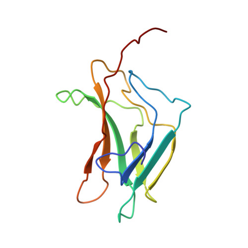
The Ig fold of the core binding factor alpha Runt domain is a member of a family of structurally and functionally related Ig-fold DNA-binding domains.
Berardi, M.J., Sun, C., Zehr, M., Abildgaard, F., Peng, J., Speck, N.A., Bushweller, J.H.(1999) Structure 7: 1247-1256
- PubMed: 10545320 Search on PubMed
- DOI: https://doi.org/10.1016/s0969-2126(00)80058-1
- Primary Citation Related Structures:
1CO1 - PubMed Abstract:
CBFA is the DNA-binding subunit of the transcription factor complex called core binding factor, or CBF. Knockout of the Cbfa2 gene in mice leads to embryonic lethality and a profound block in hematopoietic development. Chromosomal disruptions of the human CBFA gene are associated with a large percentage of human leukemias. Utilizing nuclear magnetic resonance spectroscopy we have determined the three-dimensional fold of the CBFA Runt domain in its DNA-bound state, showing that it is an s-type immunoglobulin (Ig) fold. DNA binding by the Runt domain is shown to be mediated by loop regions located at both ends of the Runt domain Ig fold. A putative site for CBFB binding has been identified; the spatial location of this site provides a rationale for the ability of CBFB to modulate the affinity of the Runt domain for DNA. Structural comparisons demonstrate that the s-type Ig fold found in the Runt domain is conserved in the Ig folds found in the DNA-binding domains of NF-kappaB, NFAT, p53, STAT-1, and the T-domain. Thus, these proteins form a family of structurally and functionally related DNA-binding domains. Unlike the other members of this family, the Runt domain utilizes loops at both ends of the Ig fold for DNA recognition.
- Department of Molecular Physiology and Biological Physics University of Virginia, Charlottesville, VA 22906, USA.
Organizational Affiliation: