1CLQ | pdb_00001clq

CRYSTAL STRUCTURE OF A REPLICATION FORK DNA POLYMERASE EDITING COMPLEX AT 2.7 A RESOLUTION


Experimental Data Snapshot

  • Method: X-RAY DIFFRACTION
  • Resolution: 2.70 Å
  • R-Value Free: 
    0.284 (Depositor), 0.276 (DCC) 
  • R-Value Work: 
    0.230 (Depositor), 0.225 (DCC) 
  • R-Value Observed: 
    0.230 (Depositor) 

Starting Model: experimental
View more details

wwPDB Validation 3D Report Full Report

Validation slider image for 1CLQ

Ligand Structure Quality Assessment 


This is version 1.3 of the entry. See complete history

Literature

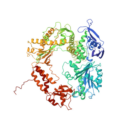
Building a replisome from interacting pieces: sliding clamp complexed to a peptide from DNA polymerase and a polymerase editing complex.

Shamoo, Y.Steitz, T.A.

(1999) Cell 99: 155-166

  • DOI: https://doi.org/10.1016/s0092-8674(00)81647-5
  • Primary Citation Related Structures: 
    1B77, 1B8H, 1CLQ

  • PubMed Abstract: 

    We have solved the crystal structures of the bacteriophage RB69 sliding clamp, its complex with a peptide essential for DNA polymerase interactions, and the DNA polymerase complexed with primer-template DNA. The editing complex structure shows a partially melted duplex DNA exiting from the exonuclease domain at an unexpected angle and significant changes in the protein structure. The clamp complex shows the C-terminal 11 residues of polymerase bound in a hydrophobic pocket, and it allows docking of the editing and clamp structures together. The peptide binds to the sliding clamp at a position identical to that of a replication inhibitor peptide bound to PCNA, suggesting that the replication inhibitor protein p21CIP1 functions by competing with eukaryotic polymerases for the same binding pocket on the clamp.


  • Organizational Affiliation
    • Department of Molecular Biophysics and Biochemistry, Yale University, New Haven, Connecticut 06520-8814, USA.

Macromolecule Content 

  • Total Structure Weight: 112.58 kDa 
  • Atom Count: 8,106 
  • Modeled Residue Count: 926 
  • Deposited Residue Count: 926 
  • Unique protein chains: 1
  • Unique nucleic acid chains: 2

Macromolecules


Find similar proteins by:|  3D Structure
Entity ID: 3
MoleculeChains  Sequence LengthOrganismDetailsImage
PROTEIN (DNA POLYMERASE)C [auth A]903Escherichia phage RB69Mutation(s): 0 
Gene Names: GENE 43
EC: 2.7.7.7 (PDB Primary Data), 3.1.11 (UniProt)
UniProt
Find proteins for Q38087 (Escherichia phage RB69)
Explore Q38087 
Go to UniProtKB:  Q38087
Entity Groups
Sequence Clusters30% Identity50% Identity70% Identity90% Identity95% Identity100% Identity
UniProt GroupQ38087
Sequence Annotations
Expand
Reference Sequence
Find similar nucleic acids by:  Sequence
Entity ID: 1
MoleculeChains LengthOrganismImage
DNA (5'-D(*GP*CP*GP*GP*AP*AP*CP*TP*AP*CP*T)-3')A [auth E]11N/A
Sequence Annotations
Expand
Reference Sequence
Find similar nucleic acids by:  Sequence
Entity ID: 2
MoleculeChains LengthOrganismImage
DNA (5'-D(*AP*GP*TP*AP*GP*TP*TP*CP*CP*GP*CP*G)-3')B [auth D]12N/A
Sequence Annotations
Expand
Reference Sequence

Small Molecules

Ligands 2 Unique
IDChains Name / Formula / InChI Key2D Diagram3D Interactions
GDP

Query on GDP


Download:Ideal Coordinates CCD File
M [auth A]GUANOSINE-5'-DIPHOSPHATE
C10 H15 N5 O11 P2
QGWNDRXFNXRZMB-UUOKFMHZSA-N
CA

Query on CA


Download:Ideal Coordinates CCD File
D [auth E]
E [auth D]
F [auth D]
G [auth A]
H [auth A]
D [auth E],
E [auth D],
F [auth D],
G [auth A],
H [auth A],
I [auth A],
J [auth A],
K [auth A],
L [auth A]
CALCIUM ION
Ca
BHPQYMZQTOCNFJ-UHFFFAOYSA-N

Experimental Data & Validation

Experimental Data

  • Method: X-RAY DIFFRACTION
  • Resolution: 2.70 Å
  • R-Value Free:  0.284 (Depositor), 0.276 (DCC) 
  • R-Value Work:  0.230 (Depositor), 0.225 (DCC) 
  • R-Value Observed: 0.230 (Depositor) 
Space Group: H 3
Unit Cell:
Length ( Å )Angle ( ˚ )
a = 209.74α = 90
b = 209.74β = 90
c = 114.25γ = 120
Software Package:
Software NamePurpose
CNSrefinement
DENZOdata reduction
SCALEPACKdata scaling
CNSphasing

Structure Validation

View Full Validation Report


Ligand Structure Quality Assessment 


Entry History 

Deposition Data

Revision History  (Full details and data files)

  • Version 1.0: 1999-10-28
    Type: Initial release
  • Version 1.1: 2008-04-26
    Changes: Version format compliance
  • Version 1.2: 2011-07-13
    Changes: Version format compliance
  • Version 1.3: 2023-08-02
    Changes: Database references, Derived calculations, Refinement description