1CJ3

MUTANT TYR38GLU OF PARA-HYDROXYBENZOATE HYDROXYLASE


Experimental Data Snapshot

  • Method: X-RAY DIFFRACTION
  • Resolution: 2.50 Å
  • R-Value Work: 0.161 

wwPDB Validation   3D Report Full Report


Ligand Structure Quality Assessment 


This is version 1.5 of the entry. See complete history


Literature

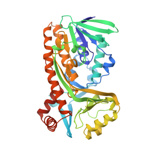
Switch of coenzyme specificity of p-hydroxybenzoate hydroxylase.

Eppink, M.H.Overkamp, K.M.Schreuder, H.A.Van Berkel, W.J.

(1999) J Mol Biol 292: 87-96

  • DOI: https://doi.org/10.1006/jmbi.1999.3015
  • Primary Citation of Related Structures:  
    1CJ2, 1CJ3, 1CJ4

  • PubMed Abstract: 

    p-Hydroxybenzoate hydroxylase (PHBH) is the archetype of the family of NAD(P)H-dependent flavoprotein aromatic hydroxylases. These enzymes share a conserved FAD-binding domain but lack a recognizable fold for binding the pyridine nucleotide. We have switched the coenzyme specificity of strictly NADPH-dependent PHBH from Pseudomonas fluorescens by site-directed mutagenesis. To that end, we altered the solvent exposed helix H2 region (residues 33-40) of the FAD-binding domain. Non-conservative selective replacements of Arg33 and Tyr38 weakened the binding of NADPH without disturbing the protein architecture. Introduction of a basic residue at position 34 increased the NADPH binding strength. Double (M2) and quadruple (M4) substitutions in the N-terminal part of helix H2 did not change the coenzyme specificity. By extending the replacements towards residues 38 and 40, M5 and M6 mutants were generated which were catalytically more efficient with NADH than with NADPH. It is concluded that specificity in P. fluorescens PHBH is conferred by interactions of Arg33, Tyr38 and Arg42 with the 2'-phosphate moiety of bound NADPH, and that introduction of an acidic group at position 38 potentially enables the recognition of the 2'-hydroxy group of NADH. This is the first report on the coenzyme reversion of a flavoprotein aromatic hydroxylase.


  • Organizational Affiliation

    Department of Biomolecular Sciences, Laboratory of Biochemistry, Wageningen University, Wageningen, 6703 HA, The Netherlands.


Macromolecules
Find similar proteins by:  (by identity cutoff)  |  3D Structure
Entity ID: 1
MoleculeChains Sequence LengthOrganismDetailsImage
PROTEIN (P-HYDROXYBENZOATE HYDROXYLASE)392Pseudomonas fluorescensMutation(s): 2 
Gene Names: POBA
EC: 1.14.13.2
UniProt
Find proteins for P00438 (Pseudomonas fluorescens)
Explore P00438 
Go to UniProtKB:  P00438
Entity Groups  
Sequence Clusters30% Identity50% Identity70% Identity90% Identity95% Identity100% Identity
UniProt GroupP00438
Sequence Annotations
Expand
  • Reference Sequence
Small Molecules
Ligands 2 Unique
IDChains Name / Formula / InChI Key2D Diagram3D Interactions
FAD
Query on FAD

Download Ideal Coordinates CCD File 
B [auth A]FLAVIN-ADENINE DINUCLEOTIDE
C27 H33 N9 O15 P2
VWWQXMAJTJZDQX-UYBVJOGSSA-N
PHB
Query on PHB

Download Ideal Coordinates CCD File 
C [auth A]P-HYDROXYBENZOIC ACID
C7 H6 O3
FJKROLUGYXJWQN-UHFFFAOYSA-N
Experimental Data & Validation

Experimental Data

  • Method: X-RAY DIFFRACTION
  • Resolution: 2.50 Å
  • R-Value Work: 0.161 
  • Space Group: C 2 2 21
Unit Cell:
Length ( Å )Angle ( ˚ )
a = 72.3α = 90
b = 146.2β = 90
c = 89.1γ = 90
Software Package:
Software NamePurpose
X-PLORmodel building
X-PLORrefinement
XDSdata reduction
XSCALEdata scaling
X-PLORphasing

Structure Validation

View Full Validation Report


Ligand Structure Quality Assessment 


Entry History 

Deposition Data

Revision History  (Full details and data files)

  • Version 1.0: 1999-04-30
    Type: Initial release
  • Version 1.1: 2007-10-16
    Changes: Version format compliance
  • Version 1.2: 2011-07-13
    Changes: Derived calculations, Version format compliance
  • Version 1.3: 2017-11-29
    Changes: Derived calculations
  • Version 1.4: 2019-11-27
    Changes: Database references
  • Version 1.5: 2023-08-09
    Changes: Data collection, Database references, Derived calculations, Refinement description