1CIZ | pdb_00001ciz

X-RAY STRUCTURE OF HUMAN STROMELYSIN CATALYTIC DOMAIN COMPLEXES WITH NON-PEPTIDE INHIBITORS: IMPLICATION FOR INHIBITOR SELECTIVITY


Experimental Data Snapshot

  • Method: X-RAY DIFFRACTION
  • Resolution: 1.64 Å
  • R-Value Free: 
    0.245 (Depositor) 
  • R-Value Work: 
    0.209 (Depositor) 

Starting Model: experimental
View more details

wwPDB Validation 3D Report Full Report

Validation slider image for 1CIZ

This is version 1.5 of the entry. See complete history

Literature

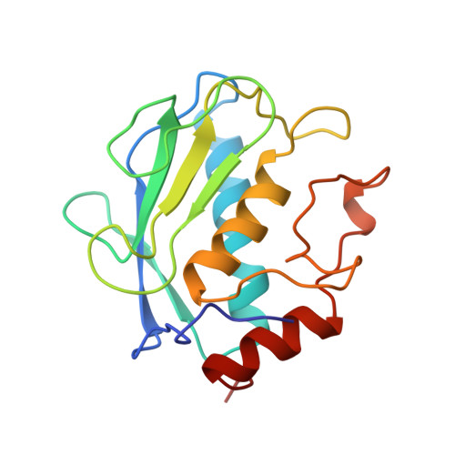
X-ray structure of human stromelysin catalytic domain complexed with nonpeptide inhibitors: implications for inhibitor selectivity.

Pavlovsky, A.G.Williams, M.G.Ye, Q.Z.Ortwine, D.F.Purchase II, C.F.White, A.D.Dhanaraj, V.Roth, B.D.Johnson, L.L.Hupe, D.Humblet, C.Blundell, T.L.

(1999) Protein Sci 8: 1455-1462

  • DOI: https://doi.org/10.1110/ps.8.7.1455
  • Primary Citation Related Structures: 
    1B8Y, 1CAQ, 1CIZ, 1QIA, 1QIC

  • PubMed Abstract: 

    Effective inhibitors of matrix metalloproteinases (MMPs), a family of connective tissue-degrading enzymes, could be useful for the treatment of diseases such as cancer, multiple sclerosis, and arthritis. Many of the known MMP inhibitors are derived from peptide substrates, with high potency in vitro but little selectivity among MMPs and poor bioavailability. We have discovered nonpeptidic MMP inhibitors with improved properties, and report here the crystal structures of human stromelysin-1 catalytic domain (SCD) complexed with four of these inhibitors. The structures were determined and refined at resolutions ranging from 1.64 to 2.0 A. Each inhibitor binds in the active site of SCD such that a bulky diphenyl piperidine moiety penetrates a deep, predominantly hydrophobic S'1 pocket. The active site structure of the SCD is similar in all four inhibitor complexes, but differs substantially from the peptide hydroxamate complex, which has a smaller side chain bound in the S'1 pocket. The largest differences occur in the loop forming the "top" of this pocket. The occupation of these nonpeptidic inhibitors in the S'1 pocket provides a structural basis to explain their selectivity among MMPs. An analysis of the unique binding mode predicts structural modifications to design improved MMP inhibitors.


  • Organizational Affiliation
    • Department of Chemistry, Parke-Davis Pharmaceutical Research, Division of Warner-Lambert Company, Ann Arbor, Michigan 48105, USA. Alexander.Pavlovsky@wl.com

Macromolecule Content 

  • Total Structure Weight: 19.74 kDa 
  • Atom Count: 1,532 
  • Modeled Residue Count: 168 
  • Deposited Residue Count: 168 
  • Unique protein chains: 1

Macromolecules

Find similar proteins by:|  3D Structure
Entity ID: 1
MoleculeChains  Sequence LengthOrganismDetailsImage
PROTEIN (STROMELYSIN-1)168Homo sapiensMutation(s): 0 
EC: 3.4.24.17
UniProt & NIH Common Fund Data Resources
Find proteins for P08254 (Homo sapiens)
Explore P08254 
Go to UniProtKB:  P08254
PHAROS:  P08254
GTEx:  ENSG00000149968 
Entity Groups
Sequence Clusters30% Identity50% Identity70% Identity90% Identity95% Identity100% Identity
UniProt GroupP08254
Sequence Annotations
Expand
Reference Sequence

Small Molecules

Ligands 4 Unique
IDChains Name / Formula / InChI Key2D Diagram3D Interactions
DPS

Query on DPS


Download:Ideal Coordinates CCD File
H [auth A]3-(1H-INDOL-3-YL)-2-[4-(4-PHENYL-PIPERIDIN-1-YL)-BENZENESULFONYLAMINO]-PROPIONIC ACID
C28 H29 N3 O4 S
ULOTXPTWJAUGGE-MHZLTWQESA-N
SO4

Query on SO4


Download:Ideal Coordinates CCD File
G [auth A]SULFATE ION
O4 S
QAOWNCQODCNURD-UHFFFAOYSA-L
ZN

Query on ZN


Download:Ideal Coordinates CCD File
B [auth A],
C [auth A]
ZINC ION
Zn
PTFCDOFLOPIGGS-UHFFFAOYSA-N
CA

Query on CA


Download:Ideal Coordinates CCD File
D [auth A],
E [auth A],
F [auth A]
CALCIUM ION
Ca
BHPQYMZQTOCNFJ-UHFFFAOYSA-N

Experimental Data & Validation

Experimental Data

  • Method: X-RAY DIFFRACTION
  • Resolution: 1.64 Å
  • R-Value Free:  0.245 (Depositor) 
  • R-Value Work:  0.209 (Depositor) 
Space Group: P 41 21 2
Unit Cell:
Length ( Å )Angle ( ˚ )
a = 69.37α = 90
b = 69.37β = 90
c = 74.4γ = 90
Software Package:
Software NamePurpose
AMoREphasing
X-PLORrefinement
DENZOdata reduction
SCALEPACKdata scaling

Structure Validation

View Full Validation Report


Entry History 

Revision History  (Full details and data files)

  • Version 1.0: 1999-09-01
    Type: Initial release
  • Version 1.1: 2007-10-16
    Changes: Version format compliance
  • Version 1.2: 2011-07-13
    Changes: Derived calculations, Version format compliance
  • Version 1.3: 2018-01-24
    Changes: Database references
  • Version 1.4: 2018-01-31
    Changes: Database references
  • Version 1.5: 2023-08-09
    Changes: Data collection, Database references, Derived calculations, Refinement description