1CGM

STRUCTURE DETERMINATION OF CUCUMBER GREEN MOTTLE MOSAIC VIRUS BY X-RAY FIBER DIFFRACTION. SIGNIFICANCE FOR THE EVOLUTION OF TOBAMOVIRUSES


Experimental Data Snapshot

  • Method: FIBER DIFFRACTION
  • Resolution: 3.40 Å
  • R-Value Work: 0.093 
  • R-Value Observed: 0.093 

wwPDB Validation   3D Report Full Report


This is version 1.3 of the entry. See complete history


Literature

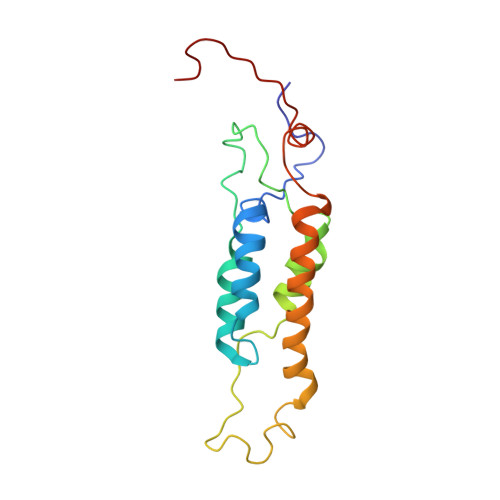
Structure determination of cucumber green mottle mosaic virus by X-ray fiber diffraction. Significance for the evolution of tobamoviruses.

Wang, H.Stubbs, G.

(1994) J Mol Biol 239: 371-384

  • DOI: https://doi.org/10.1006/jmbi.1994.1379
  • Primary Citation of Related Structures:  
    1CGM

  • PubMed Abstract: 

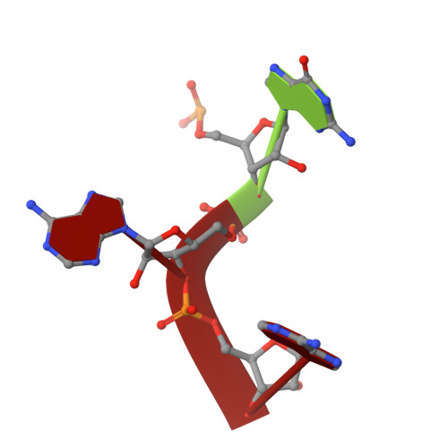
    Cucumber green mottle mosaic virus (CGMMV) is a rod-shaped virus of the tobacco mosaic virus (TMV) group. The structure of cucumber green mottle mosaic virus has been determined by fiber diffraction methods at 3.4 A resolution, and refined by molecular dynamics methods to an R factor of 0.093. Disassembly of TMV is driven by the mutual repulsion of intersubunit carboxyl-carboxylate pairs, but one of these pairs is not conserved in CGMMV. An alternative pair, located about 5 A from the site of the TMV pair, has been found in CGMMV. Comparison of the two structures suggests that the carboxylate groups are free to migrate in the subunit interfaces during evolution.


  • Organizational Affiliation

    Department of Molecular Biology, Vanderbilt University, Nashville, TN 37235.


Macromolecules

Find similar proteins by:  (by identity cutoff)  |  3D Structure
Entity ID: 2
MoleculeChains Sequence LengthOrganismDetailsImage
CUCUMBER GREEN MOTTLE MOSAIC VIRUSB [auth E]161Cucumber green mottle mosaic virusMutation(s): 0 
UniProt
Find proteins for P69475 (Cucumber green mottle mosaic virus (strain watermelon W))
Explore P69475 
Go to UniProtKB:  P69475
Entity Groups  
Sequence Clusters30% Identity50% Identity70% Identity90% Identity95% Identity100% Identity
UniProt GroupP69475
Sequence Annotations
Expand
  • Reference Sequence

Find similar nucleic acids by:  Sequence   |   3D Structure  

Entity ID: 1
MoleculeChains LengthOrganismImage
RNA (5'-R(P*GP*AP*A)-3')A [auth I]3N/A
Sequence Annotations
Expand
  • Reference Sequence
Experimental Data & Validation

Experimental Data

  • Method: FIBER DIFFRACTION
  • Resolution: 3.40 Å
  • R-Value Work: 0.093 
  • R-Value Observed: 0.093 
  • Space Group: P 1
Unit Cell:
Length ( Å )Angle ( ˚ )
a = 1α = 90
b = 1β = 90
c = 70.8γ = 90
Software Package:
Software NamePurpose
X-PLORmodel building
X-PLORrefinement
X-PLORphasing

Structure Validation

View Full Validation Report


Entry History 

Deposition Data

Revision History  (Full details and data files)

  • Version 1.0: 1994-04-30
    Type: Initial release
  • Version 1.1: 2008-03-03
    Changes: Version format compliance
  • Version 1.2: 2011-07-13
    Changes: Version format compliance
  • Version 1.3: 2017-11-29
    Changes: Derived calculations, Other