1CG1

STRUCTURE OF THE MUTANT (K16Q) OF ADENYLOSUCCINATE SYNTHETASE FROM E. COLI COMPLEXED WITH HADACIDIN, GDP, 6-PHOSPHORYL-IMP, AND MG2+


Experimental Data Snapshot

  • Method: X-RAY DIFFRACTION
  • Resolution: 2.50 Å
  • R-Value Free: 0.249 
  • R-Value Work: 0.169 

wwPDB Validation   3D Report Full Report


This is version 1.4 of the entry. See complete history


Literature

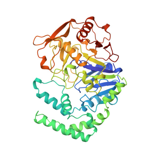
Mechanistic implications from crystalline complexes of wild-type and mutant adenylosuccinate synthetases from Escherichia coli.

Choe, J.Y.Poland, B.W.Fromm, H.J.Honzatko, R.B.

(1999) Biochemistry 38: 6953-6961

  • DOI: https://doi.org/10.1021/bi990159s
  • Primary Citation of Related Structures:  
    1CG0, 1CG1, 1CG3, 1CG4

  • PubMed Abstract: 
  • Asp13 and His41 are essential residues of adenylosuccinate synthetase, putatively catalyzing the formation of adenylosuccinate from an intermediate of 6-phosphoryl-IMP. Wild-type adenylosuccinate synthetase and three mutant synthetases (Arg143 --> Leu, Lys16 --> Gln, and Arg303 --> Leu) from Eschericha coli have been crystallized in the presence of IMP, hadacidin (an analogue of L-aspartate), Mg2+, and GTP ...

    Asp13 and His41 are essential residues of adenylosuccinate synthetase, putatively catalyzing the formation of adenylosuccinate from an intermediate of 6-phosphoryl-IMP. Wild-type adenylosuccinate synthetase and three mutant synthetases (Arg143 --> Leu, Lys16 --> Gln, and Arg303 --> Leu) from Eschericha coli have been crystallized in the presence of IMP, hadacidin (an analogue of L-aspartate), Mg2+, and GTP. The active site of each complex contains 6-phosphoryl-IMP, Mg2+, GDP, and hadacidin, except for the Arg303 --> Leu mutant, which does not bind hadacidin. In response to the formation of 6-phosphoryl-IMP, Asp13 enters the inner coordination sphere of the active site Mg2+. His41 hydrogen bonds with 6-phosphoryl-IMP, except in the Arg303 --> Leu complex, where it remains bound to the guanine nucleotide. Hence, recognition of the active site Mg2+ by Asp13 evidently occurs after the formation of 6-phosphoryl-IMP, but recognition of the intermediate by His41 may require the association of L-aspartate with the active site. Structures reported here support a mechanism in which Asp13 and His41 act as the catalytic base and acid, respectively, in the formation of 6-phosphoryl-IMP, and then act together as catalytic acids in the subsequent formation of adenylosuccinate.


    Organizational Affiliation

    Department of Biochemistry, Biophysics, and Molecular Biology, Iowa State University, Ames 50011, USA.


Macromolecules
Find similar proteins by:  (by identity cutoff)  |  3D Structure
Entity ID: 1
MoleculeChainsSequence LengthOrganismDetailsImage
PROTEIN (ADENYLOSUCCINATE SYNTHETASE)A431Escherichia coli K-12Mutation(s): 1 
Gene Names: purAadeKb4177JW4135
EC: 6.3.4.4
UniProt
Find proteins for P0A7D4 (Escherichia coli (strain K12))
Explore P0A7D4 
Go to UniProtKB:  P0A7D4
Entity Groups  
Sequence Clusters30% Identity50% Identity70% Identity90% Identity95% Identity100% Identity
UniProt GroupP0A7D4
Protein Feature View
Expand
  • Reference Sequence
Small Molecules
Ligands 4 Unique
IDChainsName / Formula / InChI Key2D Diagram3D Interactions
GDP
Query on GDP

Download Ideal Coordinates CCD File 
E [auth A]GUANOSINE-5'-DIPHOSPHATE
C10 H15 N5 O11 P2
QGWNDRXFNXRZMB-UUOKFMHZSA-N
 Ligand Interaction
IMO
Query on IMO

Download Ideal Coordinates CCD File 
D [auth A]6-O-PHOSPHORYL INOSINE MONOPHOSPHATE
C10 H14 N4 O11 P2
RXRZOKQPANIEDW-KQYNXXCUSA-N
 Ligand Interaction
HDA
Query on HDA

Download Ideal Coordinates CCD File 
C [auth A]HADACIDIN
C3 H5 N O4
URJHVPKUWOUENU-UHFFFAOYSA-N
 Ligand Interaction
MG
Query on MG

Download Ideal Coordinates CCD File 
B [auth A]MAGNESIUM ION
Mg
JLVVSXFLKOJNIY-UHFFFAOYSA-N
 Ligand Interaction
Experimental Data & Validation

Experimental Data

  • Method: X-RAY DIFFRACTION
  • Resolution: 2.50 Å
  • R-Value Free: 0.249 
  • R-Value Work: 0.169 
  • Space Group: P 32 2 1
Unit Cell:
Length ( Å )Angle ( ˚ )
a = 80.37α = 90
b = 80.37β = 90
c = 158.52γ = 120
Software Package:
Software NamePurpose
X-PLORmodel building
X-PLORrefinement
X-GENdata reduction
X-GENdata scaling
X-PLORphasing

Structure Validation

View Full Validation Report


Entry History 

Deposition Data

Revision History  (Full details and data files)

  • Version 1.0: 1999-06-17
    Type: Initial release
  • Version 1.1: 2008-04-26
    Changes: Version format compliance
  • Version 1.2: 2011-07-13
    Changes: Derived calculations, Version format compliance
  • Version 1.3: 2017-10-04
    Changes: Refinement description
  • Version 1.4: 2018-03-14
    Changes: Database references