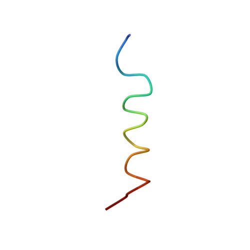
NMR solution structure of a complex of calmodulin with a binding peptide of the Ca2+ pump.
Elshorst, B., Hennig, M., Forsterling, H., Diener, A., Maurer, M., Schulte, P., Schwalbe, H., Griesinger, C., Krebs, J., Schmid, H., Vorherr, T., Carafoli, E.(1999) Biochemistry 38: 12320-12332
- PubMed: 10493800
- DOI: https://doi.org/10.1021/bi9908235
- Primary Citation of Related Structures:
1CFF - PubMed Abstract:
The three-dimensional structure of the complex between calmodulin (CaM) and a peptide corresponding to the N-terminal portion of the CaM-binding domain of the plasma membrane calcium pump, the peptide C20W, has been solved by heteronuclear three-dimensional nuclear magnetic resonance (NMR) spectroscopy. The structure calculation is based on a total of 1808 intramolecular NOEs and 49 intermolecular NOEs between the peptide C20W and calmodulin from heteronuclear-filtered NOESY spectra and a half-filtered experiment, respectively. Chemical shift differences between free Ca(2+)-saturated CaM and its complex with C20W as well as the structure calculation reveal that C20W binds solely to the C-terminal half of CaM. In addition, comparison of the methyl resonances of the nine assigned methionine residues of free Ca(2+)-saturated CaM with those of the CaM/C20W complex revealed a significant difference between the N-terminal and the C-terminal domain; i.e., resonances in the N-terminal domain of the complex were much more similar to those reported for free CaM in contrast to those in the C-terminal half which were significantly different not only from the resonances of free CaM but also from those reported for the CaM/M13 complex. As a consequence, the global structure of the CaM/C20W complex is unusual, i.e., different from other peptide calmodulin complexes, since we find no indication for a collapsed structure. The fine modulation in the peptide protein interface shows a number of differences to the CaM/M13 complex studied by Ikura et al. [Ikura, M., Clore, G. M., Gronenborn, A. M., Zhu, G., Klee, C. B., and Bax, A. (1992) Science 256, 632-638]. The unusual binding mode to only the C-terminal half of CaM is in agreement with the biochemical observation that the calcium pump can be activated by the C-terminal half of CaM alone [Guerini, D., Krebs, J., and Carafoli, E. (1984) J. Biol. Chem. 259, 15172-15177].
- Institute of Organic Chemistry, University of Frankfurt, Germany.
Organizational Affiliation:


