1CEE

SOLUTION STRUCTURE OF CDC42 IN COMPLEX WITH THE GTPASE BINDING DOMAIN OF WASP


Experimental Data Snapshot

  • Method: SOLUTION NMR
  • Conformers Calculated: 80 
  • Conformers Submitted: 20 
  • Selection Criteria: LEAST RESTRAINT VIOLATION 

wwPDB Validation   3D Report Full Report


This is version 1.4 of the entry. See complete history


Literature

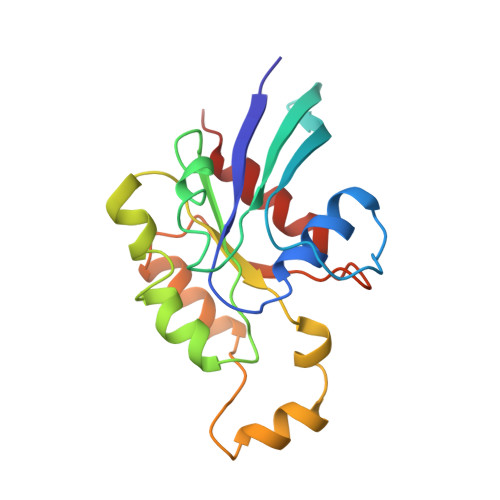
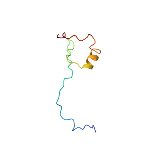
Structure of Cdc42 in complex with the GTPase-binding domain of the 'Wiskott-Aldrich syndrome' protein.

Abdul-Manan, N.Aghazadeh, B.Liu, G.A.Majumdar, A.Ouerfelli, O.Siminovitch, K.A.Rosen, M.K.

(1999) Nature 399: 379-383

  • DOI: https://doi.org/10.1038/20726
  • Primary Citation of Related Structures:  
    1CEE

  • PubMed Abstract: 

    The Rho-family GTP-hydrolysing proteins (GTPases), Cdc42, Rac and Rho, act as molecular switches in signalling pathways that regulate cytoskeletal architecture, gene expression and progression of the cell cycle. Cdc42 and Rac transmit many signals through GTP-dependent binding to effector proteins containing a Cdc42/Rac-interactive-binding (CRIB) motif. One such effector, the Wiskott-Aldrich syndrome protein (WASP), is postulated to link activation of Cdc42 directly to the rearrangement of actin. Human mutations in WASP cause severe defects in haematopoletic cell function, leading to clinical symptoms of thrombocytopenia, immunodeficiency and eczema. Here we report the solution structure of a complex between activated Cdc42 and a minimal GTPase-binding domain (GBD) from WASP. An extended amino-terminal GBD peptide that includes the CRIB motif contacts the switch I, beta2 and alpha5 regions of Cdc42. A carboxy-terminal beta-hairpin and alpha-helix pack against switch II. The Phe-X-His-X2-His portion of the CRIB motif and the alpha-helix appear to mediate sensitivity to the nucleotide switch through contacts to residues 36-40 of Cdc42. Discrimination between the Rho-family members is likely to be governed by GBD contacts to the switch I and alpha5 regions of the GTPases. Structural and biochemical data suggest that GBD-sequence divergence outside the CRIB motif may reflect additional regulatory interactions with functional domains that are specific to individual effectors.


  • Organizational Affiliation

    Cellular Biochemistry and Biophysics Program, Memorial Sloan-Kettering Cancer Center, New York, New York 10021, USA.


Macromolecules
Find similar proteins by:  (by identity cutoff)  |  3D Structure
Entity ID: 1
MoleculeChains Sequence LengthOrganismDetailsImage
GTP-BINDING RHO-LIKE PROTEIN179Homo sapiensMutation(s): 0 
UniProt & NIH Common Fund Data Resources
Find proteins for P60953 (Homo sapiens)
Explore P60953 
Go to UniProtKB:  P60953
PHAROS:  P60953
GTEx:  ENSG00000070831 
Entity Groups  
Sequence Clusters30% Identity50% Identity70% Identity90% Identity95% Identity100% Identity
UniProt GroupP60953
Sequence Annotations
Expand
  • Reference Sequence
Find similar proteins by:  (by identity cutoff)  |  3D Structure
Entity ID: 2
MoleculeChains Sequence LengthOrganismDetailsImage
WISKOTT-ALDRICH SYNDROME PROTEIN WASP59Homo sapiensMutation(s): 0 
UniProt & NIH Common Fund Data Resources
Find proteins for P42768 (Homo sapiens)
Explore P42768 
Go to UniProtKB:  P42768
PHAROS:  P42768
GTEx:  ENSG00000015285 
Entity Groups  
Sequence Clusters30% Identity50% Identity70% Identity90% Identity95% Identity100% Identity
UniProt GroupP42768
Sequence Annotations
Expand
  • Reference Sequence
Small Molecules
Ligands 2 Unique
IDChains Name / Formula / InChI Key2D Diagram3D Interactions
GCP
Query on GCP

Download Ideal Coordinates CCD File 
D [auth A]PHOSPHOMETHYLPHOSPHONIC ACID GUANYLATE ESTER
C11 H18 N5 O13 P3
PHBDHXOBFUBCJD-KQYNXXCUSA-N
MG
Query on MG

Download Ideal Coordinates CCD File 
C [auth A]MAGNESIUM ION
Mg
JLVVSXFLKOJNIY-UHFFFAOYSA-N
Experimental Data & Validation

Experimental Data

  • Method: SOLUTION NMR
  • Conformers Calculated: 80 
  • Conformers Submitted: 20 
  • Selection Criteria: LEAST RESTRAINT VIOLATION 

Structure Validation

View Full Validation Report


Entry History 

Deposition Data

Revision History  (Full details and data files)

  • Version 1.0: 1999-06-30
    Type: Initial release
  • Version 1.1: 2008-04-26
    Changes: Version format compliance
  • Version 1.2: 2011-07-13
    Changes: Version format compliance
  • Version 1.3: 2022-02-16
    Changes: Data collection, Database references, Derived calculations
  • Version 1.4: 2023-12-27
    Changes: Data collection