1CBX | pdb_00001cbx

CRYSTAL STRUCTURE OF THE COMPLEX BETWEEN CARBOXYPEPTIDASE A AND THE BIPRODUCT ANALOG INHIBITOR L-BENZYLSUCCINATE AT 2.0 ANGSTROMS RESOLUTION


Experimental Data Snapshot

  • Method: X-RAY DIFFRACTION
  • Resolution: 2.00 Å
  • R-Value Observed: 
    0.166 (Depositor) 

wwPDB Validation 3D Report Full Report

Validation slider image for 1CBX

This is version 1.4 of the entry. See complete history

Literature

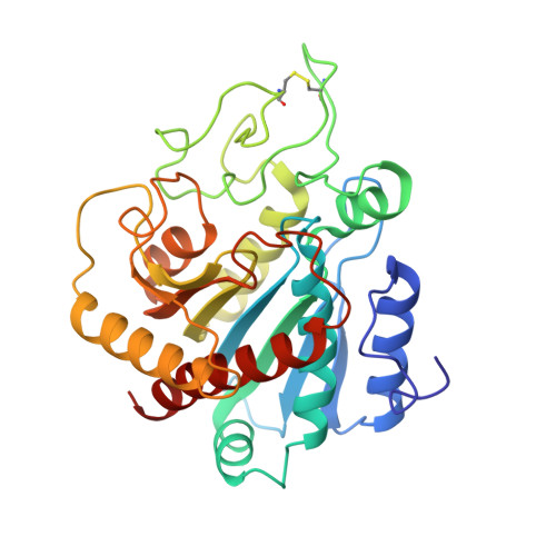
Crystal structure of the complex between carboxypeptidase A and the biproduct analog inhibitor L-benzylsuccinate at 2.0 A resolution.

Mangani, S.Carloni, P.Orioli, P.

(1992) J Mol Biology 223: 573-578

  • DOI: https://doi.org/10.1016/0022-2836(92)90671-6
  • Primary Citation Related Structures: 
    1CBX

  • PubMed Abstract: 

    The X-ray crystal structure of the carboxypeptidase A-L-benzylsuccinate complex has been refined at 2.0 A resolution to a final R-factor of 0.166. One molecule of the inhibitor binds to the enzyme active site. The terminal carboxylate forms a salt link with the guanidinium group of Arg145 and hydrogen bonds with Tyr248 and Asn144. The second carboxylate group binds to the zinc ion in an asymmetric bidentate fashion replacing the water molecule of the native structure. The zinc ion moves 0.5 A from its position in the native structure to accommodate the inhibitor binding. The overall stereochemistry around the zinc can be considered a distorted tetrahedron, although six atoms of the co-ordinated groups lie within 3.0 A from the zinc ion. The key for the strong inhibitory properties of L-benzylsuccinate can be found in its ability both to co-ordinate the zinc and to form a short carboxyl-carboxylate-type hydrogen bond (2.5 A) with Glu270.


  • Organizational Affiliation
    • Dipartimento di Chimica, Università di Siena, Italy.

Macromolecule Content 

  • Total Structure Weight: 34.72 kDa 
  • Atom Count: 2,631 
  • Modeled Residue Count: 307 
  • Deposited Residue Count: 307 
  • Unique protein chains: 1

Macromolecules

Find similar proteins by:|  3D Structure
Entity ID: 1
MoleculeChains  Sequence LengthOrganismDetailsImage
CARBOXYPEPTIDASE A307Bos taurusMutation(s): 0 
EC: 3.4.17.1
UniProt
Find proteins for P00730 (Bos taurus)
Explore P00730 
Go to UniProtKB:  P00730
Entity Groups
Sequence Clusters30% Identity50% Identity70% Identity90% Identity95% Identity100% Identity
UniProt GroupP00730
Sequence Annotations
Expand
Reference Sequence

Small Molecules

Ligands 2 Unique
IDChains Name / Formula / InChI Key2D Diagram3D Interactions
BZS

Query on BZS


Download:Ideal Coordinates CCD File
C [auth A]L-BENZYLSUCCINIC ACID
C11 H12 O4
GTOFKXZQQDSVFH-SECBINFHSA-N
ZN

Query on ZN


Download:Ideal Coordinates CCD File
B [auth A]ZINC ION
Zn
PTFCDOFLOPIGGS-UHFFFAOYSA-N

Experimental Data & Validation

Experimental Data

  • Method: X-RAY DIFFRACTION
  • Resolution: 2.00 Å
  • R-Value Observed: 0.166 (Depositor) 
Space Group: P 1 21 1
Unit Cell:
Length ( Å )Angle ( ˚ )
a = 51.6α = 90
b = 60.27β = 97.27
c = 47.25γ = 90
Software Package:
Software NamePurpose
TNTrefinement

Structure Validation

View Full Validation Report


Entry History 

Deposition Data

Revision History  (Full details and data files)

  • Version 1.0: 1994-01-31
    Type: Initial release
  • Version 1.1: 2008-03-24
    Changes: Version format compliance
  • Version 1.2: 2011-07-13
    Changes: Version format compliance
  • Version 1.3: 2017-11-29
    Changes: Derived calculations, Other
  • Version 1.4: 2024-10-30
    Changes: Data collection, Database references, Derived calculations, Structure summary