1C4R

THE STRUCTURE OF THE LIGAND-BINDING DOMAIN OF NEUREXIN 1BETA: REGULATION OF LNS DOMAIN FUNCTION BY ALTERNATIVE SPLICING


Experimental Data Snapshot

  • Method: X-RAY DIFFRACTION
  • Resolution: 2.60 Å
  • R-Value Free: 0.279 
  • R-Value Work: 0.249 
  • R-Value Observed: 0.249 

wwPDB Validation   3D Report Full Report


This is version 1.3 of the entry. See complete history


Literature

The structure of the ligand-binding domain of neurexin Ibeta: regulation of LNS domain function by alternative splicing.

Rudenko, G.Nguyen, T.Chelliah, Y.Sudhof, T.C.Deisenhofer, J.

(1999) Cell 99: 93-101

  • DOI: https://doi.org/10.1016/s0092-8674(00)80065-3
  • Primary Citation of Related Structures:  
    1C4R

  • PubMed Abstract: 

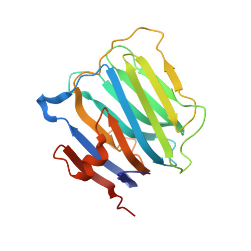
    Neurexins are expressed in hundreds of isoforms on the neuronal cell surface, where they may function as cell recognition molecules. Neurexins contain LNS domains, folding units found in many proteins like the G domain of laminin A, agrin, and slit. The crystal structure of neurexin Ibeta, a single LNS domain, reveals two seven-stranded beta sheets forming a jelly roll fold with unexpected structural similarity to lectins. The LNS domains of neurexin and agrin undergo alternative splicing that modulates their affinity for protein ligands in a neuron-specific manner. These splice sites are localized within loops at one edge of the jelly roll, suggesting a distinct protein interaction surface in LNS domains that is regulated by alternative splicing.


  • Organizational Affiliation

    Howard Hughes Medical Institute, Department of Biochemistry, University of Texas Southwestern Medical Center, Dallas 75235-9050, USA.


Macromolecules
Find similar proteins by:  (by identity cutoff)  |  3D Structure
Entity ID: 1
MoleculeChains Sequence LengthOrganismDetailsImage
NEUREXIN-I BETA
A, B, C, D, E
A, B, C, D, E, F, G, H
182Rattus norvegicusMutation(s): 0 
UniProt
Find proteins for Q63373 (Rattus norvegicus)
Explore Q63373 
Go to UniProtKB:  Q63373
Entity Groups  
Sequence Clusters30% Identity50% Identity70% Identity90% Identity95% Identity100% Identity
UniProt GroupQ63373
Sequence Annotations
Expand
  • Reference Sequence
Experimental Data & Validation

Experimental Data

  • Method: X-RAY DIFFRACTION
  • Resolution: 2.60 Å
  • R-Value Free: 0.279 
  • R-Value Work: 0.249 
  • R-Value Observed: 0.249 
  • Space Group: P 21 21 2
Unit Cell:
Length ( Å )Angle ( ˚ )
a = 116.6α = 90
b = 195.9β = 90
c = 103.61γ = 90
Software Package:
Software NamePurpose
CNSrefinement
CNSphasing

Structure Validation

View Full Validation Report


Entry History 

Deposition Data

Revision History  (Full details and data files)

  • Version 1.0: 2000-10-04
    Type: Initial release
  • Version 1.1: 2008-04-26
    Changes: Version format compliance
  • Version 1.2: 2011-07-13
    Changes: Version format compliance
  • Version 1.3: 2023-12-27
    Changes: Data collection, Database references