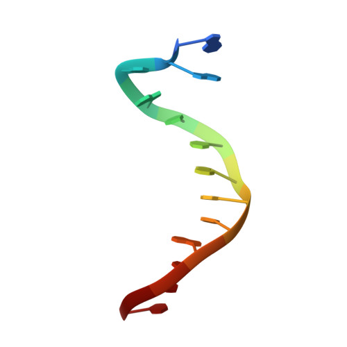
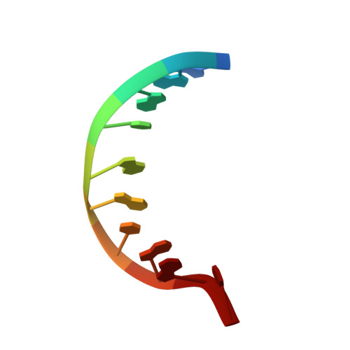
Solution structures of aminofluorene [AF]-stacked conformers of the syn [AF]-C8-dG adduct positioned opposite dC or dA at a template-primer junction.
Gu, Z., Gorin, A., Hingerty, B.E., Broyde, S., Patel, D.J.(1999) Biochemistry 38: 10855-10870
- PubMed: 10451382 Search on PubMed
- DOI: https://doi.org/10.1021/bi991266p
- Primary Citation Related Structures:
1C0Y - PubMed Abstract:
A solution structural study has been undertaken on the aminofluorene-C8-dG ([AF]dG) adduct located at a single-strand-double-strand d(A1-A2-C3-[AF]G4-C5-T6-A7-C8-C9-A10-T11-C12-C13). d(G14-G15-A16-T17-G18-G19-T20- A21-G22-N23) 13/10-mer junction (N = C or A) using proton-proton distance restraints derived from NMR data in combination with intensity-based relaxation matrix refinement computations. This single-strand-double-strand junction models one arm of a replication fork composed of a 13-mer template strand which contains the [AF]dG modification site and a 10-mer primer strand which has been elongated up to the modified guanine with either its complementary dC partner or a dA mismatch. The solution structures establish that the duplex segment retains a minimally perturbed B-DNA conformation with Watson-Crick hydrogen-bonding retained up to the dC5.dG22 base pair. The guanine ring of the [AF]dG4 adduct adopts a syn glycosidic torsion angle and is displaced into the major groove when positioned opposite dC or dA residues. This base displacement of the modified guanine is accompanied by stacking of one face of the aminofluorene ring of [AF]dG4 with the dC5.dG22 base pair, while the other face of the aminofluorene ring is stacked with the purine ring of the nonadjacent dA2 residue. By contrast, the dC and dA residues opposite the junctional [AF]dG4 adduct site adopt distinctly different alignments. The dC23 residue positioned opposite the adduct site is looped out into the minor groove by the aminofluorene ring. The syn displaced orientation of the modified dG with stacking of the aminofluorene and the looped out position of the partner dC could be envisioned to cause polymerase stalling associated with subsequent misalignment leading to frameshift mutations in appropriate sequences. The dA23 residue positioned opposite the adduct site is positioned in the major groove with its purine ring aligned face down over the van der Waals surface of the major groove and its amino group directed toward the T6.A21 base pair. The Hoogsteen edge of the modified guanine of [AF]dG4 and the Watson-Crick edge of dA23 positioned opposite it are approximately coplanar and directed toward each other but are separated by twice the hydrogen-bonding distance required for pairing. This structure of [AF]dG opposite dA at a model template-primer junctional site can be compared with a previous structure of [AF]dG opposite dA within a fully paired duplex [Norman, D., Abuaf, P., Hingerty, B. E., Live, D. , Grunberger, D., Broyde, S., and Patel, D. J. (1989) Biochemistry 28, 7462-7476]. The alignment of the Hoogsteen edge of [AF]dG (syn) positioned opposite the Watson-Crick edge of dA (anti) has been observed for both systems with the separation greater in the case of the junctional alignment in the model template-primer system. However, the aminofluorene ring is positioned in the minor groove in the fully paired duplex while it stacks over the junctional base pair in the template-primer system. This suggests that the syn [AF]dG opposite dA junctional alignment can be readily incorporated within a duplex by a translation of this entity toward the minor groove.
- Cellular Biochemistry and Biophysics Program, Memorial Sloan-Kettering Cancer Center, New York 10021, USA.
Organizational Affiliation: