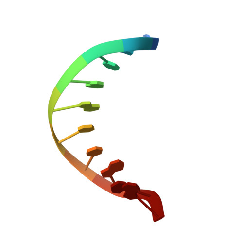
Structure determination and analysis of helix parameters in the DNA decamer d(CATGGCCATG)2 comparison of results from NMR and crystallography.
Dornberger, U., Flemming, J., Fritzsche, H.(1998) J Mol Biology 284: 1453-1463
- PubMed: 9878363 Search on PubMed
- DOI: https://doi.org/10.1006/jmbi.1998.2261
- Primary Citation Related Structures:
1BUT - PubMed Abstract:
The solution structure of the DNA decamer (CATGGCCATG)2 has been determined by NMR spectroscopy and restrained molecular dynamic and distance geometry calculations. The restrainted data set includes interproton distances and torsion angles for the deoxyribose sugar ring which were obtained by nuclear Overhauser enhancement intensities and quantitative simulation of cross-peaks from double quantum filtered correlation spectroscopy. The backbone torsion angles were constrained using experimental data from NOE cross-peaks, 1H-1H and 1H-31P-coupling constants. The NMR structure and the crystal structure of the DNA decamer deviates from the structure of the canonical form of B-DNA in a number of observable characteristics. Particularly, both structures display a specific pattern of stacking interaction in the central GGC base triplet. Furthermore, a specific local conformation of the TG/CA base-pair step is present in NMR and crystal structure, highlighting the unusually high flexibility of this DNA duplex part. The solution structure of the TG/CA base-pair step obtained by our high resolution NMR study is characterized by a positive roll angle, whereas in crystal this base-pair step tends to adopt remarkably high twist angles.
- Institut für Molekularbiologie, Friedrich-Schiller-Universität, Winzerlaer Str. 10, Jena, D-07745, Germany.
Organizational Affiliation: