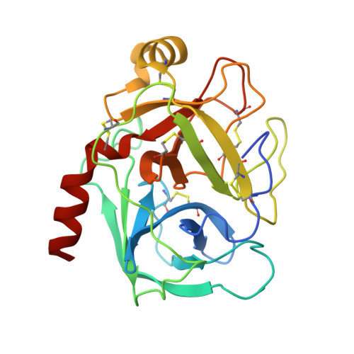
1BTP | pdb_00001btp

UNIQUE BINDING OF A NOVEL SYNTHETIC INHIBITOR, N-[3-[4-[4-(AMIDINOPHENOXY)-CARBONYL]PHENYL]-2-METHYL-2-PROPENOYL]-N-ALLYLGLYCINE METHANESULFONATE TO BOVINE TRYPSIN, REVEALED BY THE CRYSTAL STRUCTURE OF THE COMPLEX


Experimental Data Snapshot

  • Method: X-RAY DIFFRACTION
  • Resolution: 2.20 Å
  • R-Value Work: 
    0.176 (Depositor), 0.193 (DCC) 
  • R-Value Observed: 
    0.176 (Depositor) 

wwPDB Validation 3D Report Full Report

Validation slider image for 1BTP

This is version 1.4 of the entry. See complete history. 

Literature

Unique binding of a novel synthetic inhibitor, N-[3-[4-[4-(amidinophenoxy)carbonyl]phenyl]-2-methyl-2-propenoyl]- N-allylglycine methanesulfonate, to bovine trypsin, revealed by the crystal structure of the complex.

Odagaki, Y., Nakai, H., Senokuchi, K., Kawamura, M., Hamanaka, N., Nakamura, M., Tomoo, K., Ishida, T.

(1995) Biochemistry 34: 12849-12853

  • DOI: https://doi.org/10.1021/bi00039a046
  • Primary Citation Related Structures: 
    1BTP

  • PubMed Abstract: 

    Trypsin and N-[3-[4-[4-(amidinophenoxy)carbonyl]phenyl]-2-methyl-2-propenoyl]- N-allylglycine methanesulfonate (1), a newly designed and orally active synthetic trypsin inhibitor, were cocrystallized. The space group of the crystal is P2(1)2(1)2(1) with cell constants a = 63.74 A, b = 63.08 A, and c = 69.38 A, which is nearly identical to that of the orthorhombic crystal of guanidinobenzoyltrypsin. The structure was refined to a crystallographic residual R = 0.176. The refined model of the 1-trypsin complex provides the structural basis for the reaction mechanism of 1. On the basis of the present X-ray results, it is proposed that the potent inhibitory activity of 1 is mainly due to the formation of an acylated trypsin through an "inverse substrate mechanism" and its low rate of deacylation.


  • Organizational Affiliation: 
    • Minase Research Institute, Ono Pharmaceutical Co., Ltd., Osaka, Japan.

Macromolecule Content 

  • Total Structure Weight: 24.05 kDa 
  • Atom Count: 1,759 
  • Modeled Residue Count: 223 
  • Deposited Residue Count: 229 
  • Unique protein chains: 1

Macromolecules

Find similar proteins by:|  3D Structure
Entity ID: 1
MoleculeChains  Sequence LengthOrganismDetailsImage
BETA-TRYPSIN229Bos taurusMutation(s): 0 
EC: 3.4.21.4
UniProt
Find proteins for P00760 (Bos taurus)
Explore P00760 
Go to UniProtKB:  P00760
Entity Groups
Sequence Clusters30% Identity50% Identity70% Identity90% Identity95% Identity100% Identity
UniProt GroupP00760
Sequence Annotations
Expand
Reference Sequence

Small Molecules

Ligands 1 Unique
IDChains Name / Formula / InChI Key2D Diagram3D Interactions
CA

Query on CA


Download:Ideal Coordinates CCD File
B [auth A]CALCIUM ION
Ca
BHPQYMZQTOCNFJ-UHFFFAOYSA-N

Experimental Data & Validation

Experimental Data

  • Method: X-RAY DIFFRACTION
  • Resolution: 2.20 Å
  • R-Value Work:  0.176 (Depositor), 0.193 (DCC) 
  • R-Value Observed: 0.176 (Depositor) 
Space Group: P 21 21 21
Unit Cell:
Length ( Å )Angle ( ˚ )
a = 63.74α = 90
b = 63.08β = 90
c = 69.38γ = 90
Software Package:
Software NamePurpose
WISEdata collection
X-PLORmodel building
X-PLORrefinement
WISEdata reduction
X-PLORphasing

Structure Validation

View Full Validation Report


Entry History 

Deposition Data

Revision History  (Full details and data files)

  • Version 1.0: 1996-01-29
    Type: Initial release
  • Version 1.1: 2008-03-24
    Changes: Version format compliance
  • Version 1.2: 2011-07-13
    Changes: Version format compliance
  • Version 1.3: 2024-06-05
    Changes: Data collection, Database references, Derived calculations, Other
  • Version 1.4: 2024-11-06
    Changes: Structure summary