1BSU | pdb_00001bsu

STRUCTURAL AND ENERGETIC ORIGINS OF INDIRECT READOUT IN SITE-SPECIFIC DNA CLEAVAGE BY A RESTRICTION ENDONUCLEASE


Experimental Data Snapshot

  • Method: X-RAY DIFFRACTION
  • Resolution: 2.00 Å
  • R-Value Free: 
    0.258 (Depositor) 
  • R-Value Work: 
    0.183 (Depositor) 
  • R-Value Observed: 
    0.183 (Depositor) 

Starting Model: experimental
View more details

wwPDB Validation 3D Report Full Report

Validation slider image for 1BSU

This is version 1.4 of the entry. See complete history

Literature

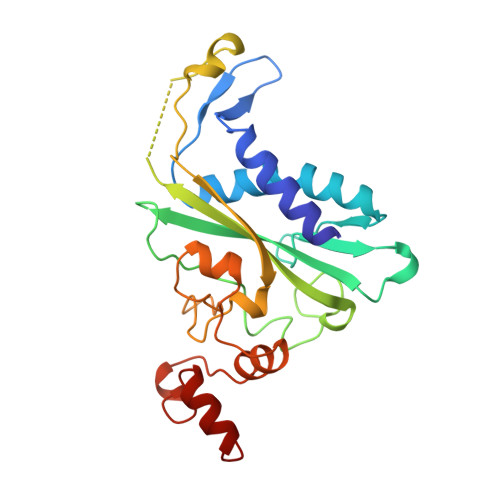
Structural and energetic origins of indirect readout in site-specific DNA cleavage by a restriction endonuclease.

Martin, A.M.Sam, M.D.Reich, N.O.Perona, J.J.

(1999) Nat Struct Biol 6: 269-277

  • DOI: https://doi.org/10.1038/6707
  • Primary Citation Related Structures: 
    1BSU, 1BUA

  • PubMed Abstract: 

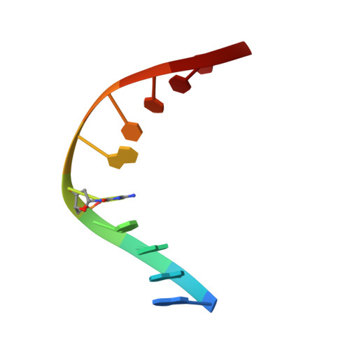
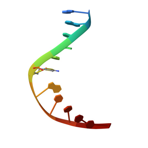
    Specific recognition by EcoRV endonuclease of its cognate, sharply bent GATATC site at the center TA step occurs solely via hydrophobic interaction with thymine methyl groups. Mechanistic kinetic analyses of base analog-substituted DNAs at this position reveal that direct readout provides 5 kcal mol(-1) toward specificity, with an additional 6-10 kcal mol(-1) arising from indirect readout. Crystal structures of several base analog complexes show that the major-groove hydrophobic contacts are crucial to forming required divalent metal-binding sites, and that indirect readout operates in part through the sequence-dependent free-energy cost of unstacking the center base-pair step of the DNA.


  • Organizational Affiliation
    • Department of Chemistry, University of California at Santa Barbara, 93106-9510, USA.

Macromolecule Content 

  • Total Structure Weight: 63.6 kDa 
  • Atom Count: 4,281 
  • Modeled Residue Count: 486 
  • Deposited Residue Count: 509 
  • Unique protein chains: 1
  • Unique nucleic acid chains: 2

Macromolecules


Find similar proteins by:|  3D Structure
Entity ID: 3
MoleculeChains  Sequence LengthOrganismDetailsImage
ENDONUCLEASE ECORV (3.1.21.4)C [auth A],
D [auth B]
244Escherichia coliMutation(s): 0 
EC: 3.1.21.4
UniProt
Find proteins for P04390 (Escherichia coli)
Explore P04390 
Go to UniProtKB:  P04390
Entity Groups
Sequence Clusters30% Identity50% Identity70% Identity90% Identity95% Identity100% Identity
UniProt GroupP04390
Sequence Annotations
Expand
Reference Sequence
Find similar nucleic acids by:  Sequence
Entity ID: 1
MoleculeChains LengthOrganismImage
DNA (5'-D(P*AP*AP*GP*AP*(5CM)P*IP*TP*CP*TP*T)-3')A [auth C]10N/A
Sequence Annotations
Expand
Reference Sequence
Find similar nucleic acids by:  Sequence
Entity ID: 2
MoleculeChains LengthOrganismImage
DNA (5'-D(*AP*AP*AP*GP*AP*(5CM)P*IP*TP*CP*TP*T)-3')B [auth D]11N/A
Sequence Annotations
Expand
Reference Sequence

Small Molecules

Experimental Data & Validation

Experimental Data

  • Method: X-RAY DIFFRACTION
  • Resolution: 2.00 Å
  • R-Value Free:  0.258 (Depositor) 
  • R-Value Work:  0.183 (Depositor) 
  • R-Value Observed: 0.183 (Depositor) 
Space Group: P 1
Unit Cell:
Length ( Å )Angle ( ˚ )
a = 50.3α = 109.1
b = 64β = 108.1
c = 49.1γ = 96.5
Software Package:
Software NamePurpose
X-PLORmodel building
X-PLORrefinement
R-AXISdata reduction
R-AXISdata scaling
X-PLORphasing

Structure Validation

View Full Validation Report


Entry History 

Deposition Data

Revision History  (Full details and data files)

  • Version 1.0: 1998-09-09
    Type: Initial release
  • Version 1.1: 2008-05-22
    Changes: Version format compliance
  • Version 1.2: 2011-07-13
    Changes: Version format compliance
  • Version 1.3: 2019-11-20
    Changes: Derived calculations
  • Version 1.4: 2023-08-02
    Changes: Database references, Derived calculations, Refinement description