1BSS

ECORV-T93A/DNA/CA2+


Experimental Data Snapshot

  • Method: X-RAY DIFFRACTION
  • Resolution: 2.15 Å
  • R-Value Free: 0.246 
  • R-Value Work: 0.184 
  • R-Value Observed: 0.184 

wwPDB Validation   3D Report Full Report


This is version 1.5 of the entry. See complete history


Literature

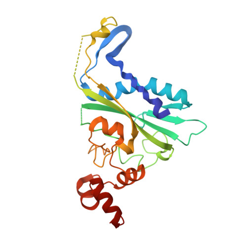
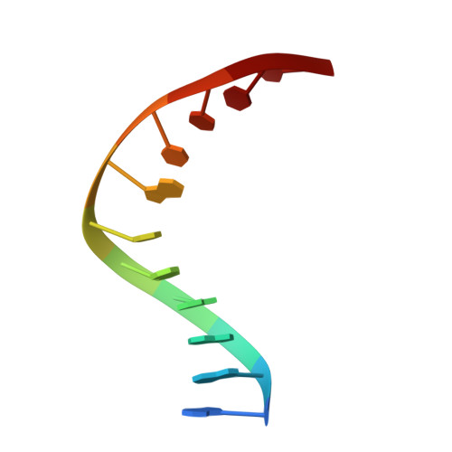
Metal ion-mediated substrate-assisted catalysis in type II restriction endonucleases

Horton, N.C.Newberry, K.J.Perona, J.J.

(1998) Proc Natl Acad Sci U S A 95: 13489-13494

  • DOI: https://doi.org/10.1073/pnas.95.23.13489
  • Primary Citation of Related Structures:  
    1BSS

  • PubMed Abstract: 

    The 2.15-A resolution cocrystal structure of EcoRV endonuclease mutant T93A complexed with DNA and Ca2+ ions reveals two divalent metals bound in one of the active sites. One of these metals is ligated through an inner-sphere water molecule to the phosphate group located 3' to the scissile phosphate. A second inner-sphere water on this metal is positioned approximately in-line for attack on the scissile phosphate. This structure corroborates the observation that the pro-SP phosphoryl oxygen on the adjacent 3' phosphate cannot be modified without severe loss of catalytic efficiency. The structural equivalence of key groups, conserved in the active sites of EcoRV, EcoRI, PvuII, and BamHI endonucleases, suggests that ligation of a catalytic divalent metal ion to this phosphate may occur in many type II restriction enzymes. Together with previous cocrystal structures, these data allow construction of a detailed model for the pretransition state configuration in EcoRV. This model features three divalent metal ions per active site and invokes assistance in the bond-making step by a conserved lysine, which stabilizes the attacking hydroxide ion nucleophile.


  • Organizational Affiliation

    Department of Chemistry and Interdepartmental Program in Biochemistry and Molecular Biology, University of California, Santa Barbara, CA 93106-9510, USA.


Macromolecules

Find similar proteins by:  (by identity cutoff)  |  3D Structure
Entity ID: 2
MoleculeChains Sequence LengthOrganismDetailsImage
ECORV ENDONUCLEASEC [auth A],
D [auth B]
244Escherichia coliMutation(s): 1 
EC: 3.1.21.4
UniProt
Find proteins for P04390 (Escherichia coli)
Explore P04390 
Go to UniProtKB:  P04390
Entity Groups  
Sequence Clusters30% Identity50% Identity70% Identity90% Identity95% Identity100% Identity
UniProt GroupP04390
Sequence Annotations
Expand
  • Reference Sequence

Find similar nucleic acids by:  Sequence   |   3D Structure  

Entity ID: 1
MoleculeChains LengthOrganismImage
5'-D(*AP*AP*AP*GP*AP*TP*AP*TP*CP*TP*T)-3'A [auth C],
B [auth D]
11N/A
Sequence Annotations
Expand
  • Reference Sequence
Small Molecules
Ligands 1 Unique
IDChains Name / Formula / InChI Key2D Diagram3D Interactions
CA
Query on CA

Download Ideal Coordinates CCD File 
E [auth A],
F [auth A]
CALCIUM ION
Ca
BHPQYMZQTOCNFJ-UHFFFAOYSA-N
Experimental Data & Validation

Experimental Data

  • Method: X-RAY DIFFRACTION
  • Resolution: 2.15 Å
  • R-Value Free: 0.246 
  • R-Value Work: 0.184 
  • R-Value Observed: 0.184 
  • Space Group: P 1
Unit Cell:
Length ( Å )Angle ( ˚ )
a = 50α = 109.2
b = 64.4β = 108.7
c = 49.2γ = 95.8
Software Package:
Software NamePurpose
X-PLORmodel building
X-PLORrefinement
X-PLORphasing

Structure Validation

View Full Validation Report


Entry History 

Deposition Data

Revision History  (Full details and data files)

  • Version 1.0: 1998-09-02
    Type: Initial release
  • Version 1.1: 2008-04-27
    Changes: Version format compliance
  • Version 1.2: 2011-07-13
    Changes: Version format compliance
  • Version 1.3: 2017-10-04
    Changes: Refinement description
  • Version 1.4: 2021-11-03
    Changes: Database references, Derived calculations
  • Version 1.5: 2023-08-02
    Changes: Refinement description