Manipulating the coordination mumber of the ferric iron within the cambialistic superoxide dismutase of Propionibacterium shermanii by changing the pH-value A crystallographic analysis
Schmidt, M.(1999) Eur J Biochem 262: 117-127
- PubMed: 10231372 Search on PubMed
- DOI: https://doi.org/10.1046/j.1432-1327.1999.00359.x
- Primary Citation Related Structures:
1BS3, 1BSM, 1BT8 - PubMed Abstract:
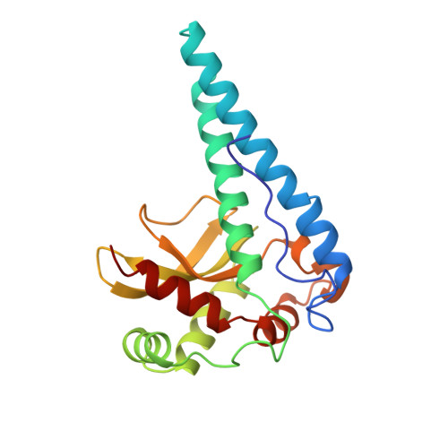
The structure of the Propionibacterium freudenreichii subspecies shermanii superoxide dismutase (SOD) was determined at various pH values. As a comparison, the structure of the fluoride coordinated SOD was solved. The SOD crystallizes at pH 6.1 in the space group C2221 with two subunits, A and B, in the asymmetric unit. An increase of the pH value changes the cell parameters slightly but not the symmetry of the crystals. The overall structure of the SOD remains a compact tetrameter and is comparable to that at pH 6.1 no matter whether the pH increases or fluoride is added. At values above pH 7.4, an additional hydroxide ion can bind to the active center. Its position is similar to the binding site of the fluoride. The coordination number changes from five to six if the pH increases or fluoride is added. The binding behavior of the hydroxide ion is different for subunit A and B. Structures at different pH-values are comparable with models derived by spectroscopic methods. The influence of temperature on the binding properties of the hydroxide ion was investigated using analysis of an X-ray structure solved at pH 8.1 and 140 K. Compared to the structure at room temperature, the structural changes are observable but remain small. The consequences of hydroxide binding to the iron are discussed.
- Physikdepartment E17, Technische Universität München, James Franck Strasse, Garching, Germany. marius@vishnu.uchicago.edu
Organizational Affiliation: