1BR9 | pdb_00001br9

HUMAN TISSUE INHIBITOR OF METALLOPROTEINASE-2


Experimental Data Snapshot

  • Method: X-RAY DIFFRACTION
  • Resolution: 2.10 Å
  • R-Value Free: 
    0.271 (Depositor) 
  • R-Value Work: 
    0.238 (Depositor), 0.224 (DCC) 

Starting Model: experimental
View more details

wwPDB Validation 3D Report Full Report

Validation slider image for 1BR9

This is version 1.4 of the entry. See complete history

Literature

Three-dimensional structure of human tissue inhibitor of metalloproteinases-2 at 2.1 A resolution.

Tuuttila, A.Morgunova, E.Bergmann, U.Lindqvist, Y.Maskos, K.Fernandez-Catalan, C.Bode, W.Tryggvason, K.Schneider, G.

(1998) J Mol Biology 284: 1133-1140

  • DOI: https://doi.org/10.1006/jmbi.1998.2223
  • Primary Citation Related Structures: 
    1BR9

  • PubMed Abstract: 

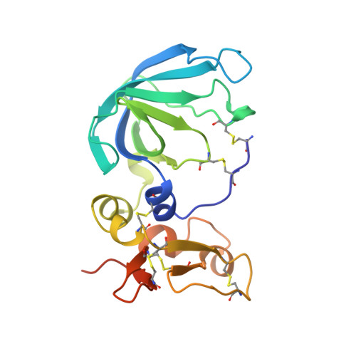
    The three-dimensional structure of human tissue inhibitor of metalloproteinases-2 (TIMP-2) was determined by X-ray crystallography to 2.1 A resolution. The structure of the inhibitor consists of two domains. The N-terminal domain (residues 1-110) is folded into a beta-barrel, similar to the oligonucleotide/oligosaccharide binding fold otherwise found in certain DNA-binding proteins. The C-terminal domain (residues 111-194) contains a parallel stranded beta-hairpin plus a beta-loop-beta motif. Comparison of the structure of uncomplexed human TIMP-2 with that of bovine TIMP-2 bound to the catalytic domain of human MMP-14 suggests an internal rotation between the two domains of approximately 13 degrees upon binding to the protease. Furthermore, local conformational differences in the two structures that might be induced by formation of the protease-inhibitor complex have been found. The most prominent of these involves residues 27-40 of the A-B beta-hairpin loop. Structure-based alignment of amino acid sequences of representatives of the TIMP family maps the sequence differences mainly to loop regions, and some of these differences are proposed to be responsible for the particular properties of the various TIMP species.


  • Organizational Affiliation
    • Department of Medical Biochemistry and Biophysics Division of Matrix Biology, Karolinska Institutet, Stockholm, S-171 77, Sweden.

Macromolecule Content 

  • Total Structure Weight: 21.78 kDa 
  • Atom Count: 1,482 
  • Modeled Residue Count: 182 
  • Deposited Residue Count: 194 
  • Unique protein chains: 1

Macromolecules

Find similar proteins by:|  3D Structure
Entity ID: 1
MoleculeChains  Sequence LengthOrganismDetailsImage
METALLOPROTEINASE-2 INHIBITOR194Homo sapiensMutation(s): 0 
UniProt & NIH Common Fund Data Resources
Find proteins for P16035 (Homo sapiens)
Explore P16035 
Go to UniProtKB:  P16035
PHAROS:  P16035
GTEx:  ENSG00000035862 
Entity Groups
Sequence Clusters30% Identity50% Identity70% Identity90% Identity95% Identity100% Identity
UniProt GroupP16035
Sequence Annotations
Expand
Reference Sequence

Experimental Data & Validation

Experimental Data

  • Method: X-RAY DIFFRACTION
  • Resolution: 2.10 Å
  • R-Value Free:  0.271 (Depositor) 
  • R-Value Work:  0.238 (Depositor), 0.224 (DCC) 
Space Group: P 21 21 2
Unit Cell:
Length ( Å )Angle ( ˚ )
a = 72.05α = 90
b = 52.7β = 90
c = 46.1γ = 90
Software Package:
Software NamePurpose
DENZOdata reduction
SCALEPACKdata scaling
AMoREphasing
REFMACrefinement

Structure Validation

View Full Validation Report


Entry History 

Deposition Data

Revision History  (Full details and data files)

  • Version 1.0: 1999-05-04
    Type: Initial release
  • Version 1.1: 2008-03-24
    Changes: Version format compliance
  • Version 1.2: 2011-07-13
    Changes: Version format compliance
  • Version 1.3: 2024-04-03
    Changes: Data collection, Database references, Refinement description
  • Version 1.4: 2024-10-16
    Changes: Structure summary