1BND | pdb_00001bnd

STRUCTURE OF THE BRAIN-DERIVED NEUROTROPHIC FACTOR(SLASH)NEUROTROPHIN 3 HETERODIMER


Experimental Data Snapshot

  • Method: X-RAY DIFFRACTION
  • Resolution: 2.30 Å
  • R-Value Work: 
    0.182 (Depositor) 
  • R-Value Observed: 
    0.182 (Depositor) 

wwPDB Validation 3D Report Full Report

Validation slider image for 1BND

This is version 1.3 of the entry. See complete history

Literature

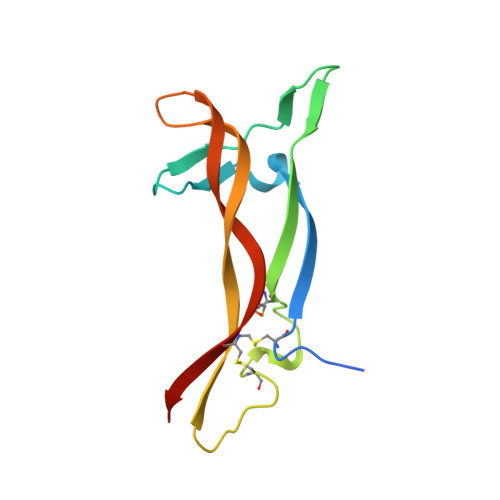
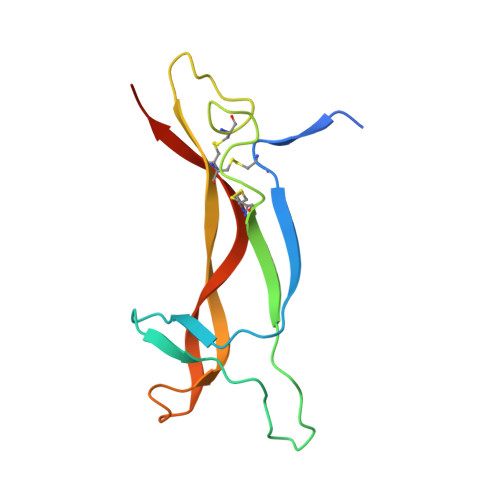
Structure of the brain-derived neurotrophic factor/neurotrophin 3 heterodimer.

Robinson, R.C.Radziejewski, C.Stuart, D.I.Jones, E.Y.

(1995) Biochemistry 34: 4139-4146

  • DOI: https://doi.org/10.1021/bi00013a001
  • Primary Citation Related Structures: 
    1BND

  • PubMed Abstract: 

    The development and sustenance of specific neuronal populations in the peripheral and central nervous systems are controlled through the binding of neurotrophic factors to high-affinity cell surface receptors. The neurotrophins (nerve growth factor, NGF; brain-derived neurotrophic factor, BDNF; neurotrophin 3, NT3; and neurotrophin 4, NT4) are dimeric molecules which share approximately 50% sequence identity. The crystal structure of the murine NGF homodimer [McDonald et al. (1991) Nature 354, 411-414] indicated that the dimer interface corresponds to regions of high sequence conservation throughout the neurotrophin family. This potential compatibility was duly exploited for the production in vitro of noncovalent heterodimers between the different neurotrophins [Radziejewski, C., & Robinson, R.C. (1993) Biochemistry 32, 13350-13356; Jungbluth et al. (1994) Eur. J. Biochem. 221, 677-685]. Here, we report the X-ray structure at 2.3 A resolution of one such heterodimer, between human BDNF, and human NT3. The NGF, BDNF, and NT3 protomers share the same topology and are structurally equivalent in regions which contribute to the dimer interface in line with the propensity of the neurotrophins to form heterodimers. Analysis of the structure of regions of the BDNF/NT3 heterodimer involved in receptor specificity led us to conclude that heterodimer binding to p75 involves distant binding sites separately located on each protomer of the heterodimer. In contrast, heterodimer interactions with the trk receptors probably utilize hybrid binding sites comprised of residues contributed by both protomers in the heterodimer. The existence of such hybrid binding sites for the trk receptor provides an explanation for the lower activity of the BDNF/NT3 heterodimer in comparison to the homodimers.(ABSTRACT TRUNCATED AT 250 WORDS)


  • Organizational Affiliation
    • Laboratory of Molecular Biophysics, Oxford University, United Kingdom.

Macromolecule Content 

  • Total Structure Weight: 27.25 kDa 
  • Atom Count: 1,796 
  • Modeled Residue Count: 217 
  • Deposited Residue Count: 238 
  • Unique protein chains: 2

Macromolecules

Find similar proteins by:|  3D Structure
Entity ID: 1
MoleculeChains  Sequence LengthOrganismDetailsImage
BRAIN DERIVED NEUROTROPHIC FACTOR119Homo sapiensMutation(s): 0 
UniProt & NIH Common Fund Data Resources
Find proteins for P23560 (Homo sapiens)
Explore P23560 
Go to UniProtKB:  P23560
PHAROS:  P23560
GTEx:  ENSG00000176697 
Entity Groups
Sequence Clusters30% Identity50% Identity70% Identity90% Identity95% Identity100% Identity
UniProt GroupP23560
Sequence Annotations
Expand
Reference Sequence
Find similar proteins by:|  3D Structure
Entity ID: 2
MoleculeChains  Sequence LengthOrganismDetailsImage
NEUROTROPHIN 3119Homo sapiensMutation(s): 0 
UniProt & NIH Common Fund Data Resources
Find proteins for P20783 (Homo sapiens)
Explore P20783 
Go to UniProtKB:  P20783
PHAROS:  P20783
GTEx:  ENSG00000185652 
Entity Groups
Sequence Clusters30% Identity50% Identity70% Identity90% Identity95% Identity100% Identity
UniProt GroupP20783
Sequence Annotations
Expand
Reference Sequence

Small Molecules

Ligands 1 Unique
IDChains Name / Formula / InChI Key2D Diagram3D Interactions
IPA

Query on IPA


Download:Ideal Coordinates CCD File
C [auth A]ISOPROPYL ALCOHOL
C3 H8 O
KFZMGEQAYNKOFK-UHFFFAOYSA-N

Experimental Data & Validation

Experimental Data

  • Method: X-RAY DIFFRACTION
  • Resolution: 2.30 Å
  • R-Value Work:  0.182 (Depositor) 
  • R-Value Observed: 0.182 (Depositor) 
Space Group: C 1 2 1
Unit Cell:
Length ( Å )Angle ( ˚ )
a = 98.4α = 90
b = 45.1β = 118.3
c = 68γ = 90
Software Package:
Software NamePurpose
DENZOdata reduction
X-PLORmodel building
X-PLORrefinement
X-PLORphasing

Structure Validation

View Full Validation Report


Entry History 

Deposition Data

Revision History  (Full details and data files)

  • Version 1.0: 1996-04-04
    Type: Initial release
  • Version 1.1: 2008-03-24
    Changes: Version format compliance
  • Version 1.2: 2011-07-13
    Changes: Derived calculations, Version format compliance
  • Version 1.3: 2024-10-30
    Changes: Data collection, Database references, Derived calculations, Structure summary