1BN8 | pdb_00001bn8

BACILLUS SUBTILIS PECTATE LYASE


Experimental Data Snapshot

  • Method: X-RAY DIFFRACTION
  • Resolution: 1.80 Å
  • R-Value Work: 
    0.179 (Depositor), 0.163 (DCC) 

wwPDB Validation 3D Report Full Report

Validation slider image for 1BN8

This is version 1.3 of the entry. See complete history

Literature

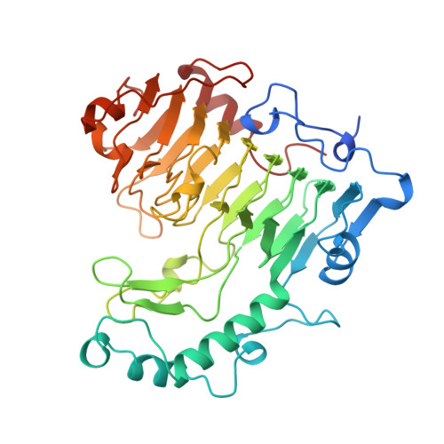
The structure of Bacillus subtilis pectate lyase in complex with calcium.

Pickersgill, R.Jenkins, J.Harris, G.Nasser, W.Robert-Baudouy, J.

(1994) Nat Struct Biol 1: 717-723

  • DOI: https://doi.org/10.1038/nsb1094-717
  • Primary Citation Related Structures: 
    1BN8

  • PubMed Abstract: 

    We have solved the structure of the Bacillus subtilis pectate lyase (BsPel) in complex with calcium. The structure consists of a parallel beta-helix domain and a loop region. The alpha L-bounded beta-strand seen in BsPel is a new element of protein structure and its frequent occurrence suggests it is an important characteristic of the parallel beta-helix. A pronounced cleft is formed between the loops and the parallel beta-helix domain and we propose that this is the active site cleft. Calcium, essential for the activity of the enzyme, binds at the bottom of this cleft and an arginine residue close to the calcium, which is conserved across all pectin and pectate lyases, may be involved in catalysis.


  • Organizational Affiliation
    • Department of Protein Engineering, Institute of Food Research, Reading, UK.

Macromolecule Content 

  • Total Structure Weight: 45.59 kDa 
  • Atom Count: 3,276 
  • Modeled Residue Count: 399 
  • Deposited Residue Count: 420 
  • Unique protein chains: 1

Macromolecules

Find similar proteins by:|  3D Structure
Entity ID: 1
MoleculeChains  Sequence LengthOrganismDetailsImage
PROTEIN (PECTATE LYASE)420Bacillus subtilisMutation(s): 0 
EC: 4.2.2.2
UniProt
Find proteins for P39116 (Bacillus subtilis (strain 168))
Explore P39116 
Go to UniProtKB:  P39116
Entity Groups
Sequence Clusters30% Identity50% Identity70% Identity90% Identity95% Identity100% Identity
UniProt GroupP39116
Sequence Annotations
Expand
Reference Sequence

Small Molecules

Ligands 1 Unique
IDChains Name / Formula / InChI Key2D Diagram3D Interactions
CA

Query on CA


Download:Ideal Coordinates CCD File
B [auth A]CALCIUM ION
Ca
BHPQYMZQTOCNFJ-UHFFFAOYSA-N

Experimental Data & Validation

Experimental Data

  • Method: X-RAY DIFFRACTION
  • Resolution: 1.80 Å
  • R-Value Work:  0.179 (Depositor), 0.163 (DCC) 
Space Group: P 1 21 1
Unit Cell:
Length ( Å )Angle ( ˚ )
a = 55.9α = 90
b = 89β = 108.93
c = 50.7γ = 90
Software Package:
Software NamePurpose
REFMACrefinement
DENZOdata reduction
SCALEPACKdata scaling

Structure Validation

View Full Validation Report


Entry History 

Deposition Data

Revision History  (Full details and data files)

  • Version 1.0: 1998-08-05
    Type: Initial release
  • Version 1.1: 2007-10-16
    Changes: Version format compliance
  • Version 1.2: 2011-07-13
    Changes: Version format compliance
  • Version 1.3: 2024-02-07
    Changes: Data collection, Database references, Derived calculations