1BJT | pdb_00001bjt

TOPOISOMERASE II RESIDUES 409-1201


Experimental Data Snapshot

  • Method: X-RAY DIFFRACTION
  • Resolution: 2.50 Å
  • R-Value Free: 
    0.269 (Depositor) 
  • R-Value Work: 
    0.227 (Depositor), 0.236 (DCC) 
  • R-Value Observed: 
    0.227 (Depositor) 

wwPDB Validation 3D Report Full Report

Validation slider image for 1BJT

This is version 1.3 of the entry. See complete history

Literature

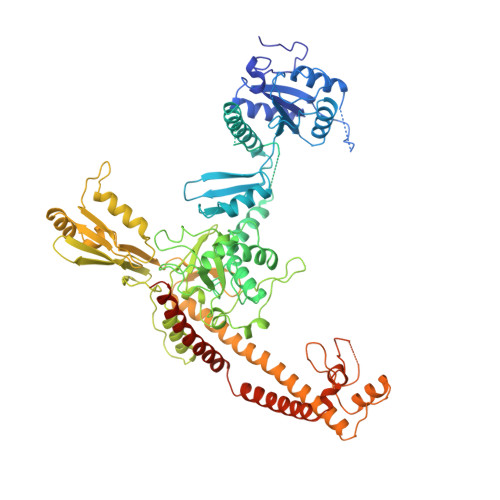
Quaternary changes in topoisomerase II may direct orthogonal movement of two DNA strands.

Fass, D.Bogden, C.E.Berger, J.M.

(1999) Nat Struct Biol 6: 322-326

  • DOI: https://doi.org/10.1038/7556
  • Primary Citation Related Structures: 
    1BJT

  • PubMed Abstract: 

    Type II DNA topoisomerases mediate the passage of one DNA duplex through a transient break in another, an event essential for chromosome segregation and cell viability. The active sites of the type II topoisomerase dimer associate covalently with the DNA break-points and must separate by at least the width of the second DNA duplex to accommodate transport. A new structure of the Saccharomyces cerevisiae topoisomerase II DNA-binding and cleavage core suggests that in addition to conformational changes in the DNA-opening platform, a dramatic reorganization of accessory domains may occur during catalysis. These conformational differences have implications for both the DNA-breaking and duplex-transport events in the topo II reaction mechanism, suggest a mechanism by which two distinct drug-resistance loci interact, and illustrate the scope of structural changes in the cycling of molecular machines.


  • Organizational Affiliation
    • Whitehead Institute for Biomedical Research, Cambridge, Massachusetts 02140, USA.

Macromolecule Content 

  • Total Structure Weight: 91.81 kDa 
  • Atom Count: 5,815 
  • Modeled Residue Count: 679 
  • Deposited Residue Count: 793 
  • Unique protein chains: 1

Macromolecules

Find similar proteins by:|  3D Structure
Entity ID: 1
MoleculeChains  Sequence LengthOrganismDetailsImage
TOPOISOMERASE II793Saccharomyces cerevisiaeMutation(s): 0 
EC: 5.99.1.3 (PDB Primary Data), 5.6.2.2 (UniProt)
UniProt
Find proteins for P06786 (Saccharomyces cerevisiae (strain ATCC 204508 / S288c))
Explore P06786 
Go to UniProtKB:  P06786
Entity Groups
Sequence Clusters30% Identity50% Identity70% Identity90% Identity95% Identity100% Identity
UniProt GroupP06786
Sequence Annotations
Expand
Reference Sequence

Experimental Data & Validation

Experimental Data

  • Method: X-RAY DIFFRACTION
  • Resolution: 2.50 Å
  • R-Value Free:  0.269 (Depositor) 
  • R-Value Work:  0.227 (Depositor), 0.236 (DCC) 
  • R-Value Observed: 0.227 (Depositor) 
Space Group: C 2 2 21
Unit Cell:
Length ( Å )Angle ( ˚ )
a = 103.4α = 90
b = 145.7β = 90
c = 161.3γ = 90
Software Package:
Software NamePurpose
X-PLORmodel building
X-PLORrefinement
DENZOdata reduction
SCALEPACKdata scaling
X-PLORphasing

Structure Validation

View Full Validation Report


Entry History 

Deposition Data

Revision History  (Full details and data files)

  • Version 1.0: 1999-05-04
    Type: Initial release
  • Version 1.1: 2008-03-24
    Changes: Version format compliance
  • Version 1.2: 2011-07-13
    Changes: Derived calculations, Version format compliance
  • Version 1.3: 2024-02-07
    Changes: Data collection, Database references, Other