1BJ4 | pdb_00001bj4

RECOMBINANT SERINE HYDROXYMETHYLTRANSFERASE (HUMAN)


Experimental Data Snapshot

  • Method: X-RAY DIFFRACTION
  • Resolution: 2.65 Å
  • R-Value Free: 
    0.226 (Depositor) 
  • R-Value Work: 
    0.210 (Depositor) 
  • R-Value Observed: 
    0.210 (Depositor) 

wwPDB Validation 3D Report Full Report

Validation slider image for 1BJ4

This is version 2.0 of the entry. See complete history

Literature

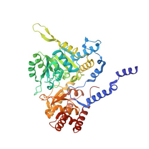
The crystal structure of human cytosolic serine hydroxymethyltransferase: a target for cancer chemotherapy

Renwick, S.B.Snell, K.Baumann, U.

(1998) Structure 6: 1105-1116

  • DOI: https://doi.org/10.1016/s0969-2126(98)00112-9
  • Primary Citation Related Structures: 
    1BJ4

  • PubMed Abstract: 

    Serine hydroxymethyltransferase (SHMT) is a ubiquitous enzyme found in all prokaryotes and eukaryotes. As an enzyme of the thymidylate synthase metabolic cycle, SHMT catalyses the retro-aldol cleavage of serine to glycine, with the resulting hydroxymethyl group being transferred to tetrahydrofolate to form 5, 10-methylene-tetrahydrofolate. The latter is the major source of one-carbon units in metabolism. Elevated SHMT activity has been shown to be coupled to the increased demand for DNA synthesis in rapidly proliferating cells, particularly tumour cells. Consequently, the central role of SHMT in nucleotide biosynthesis makes it an attractive target for cancer chemotherapy. We have solved the crystal structure of human cytosolic SHMT by multiple isomorphous replacement to 2.65 A resolution. The monomer has a fold typical for alpha class pyridoxal 5'-phosphate (PLP) dependent enzymes. The tetramer association is best described as a 'dimer of dimers' where residues from both subunits of one 'tight' dimer contribute to the active site. The crystal structure shows the evolutionary relationship between SHMT and other alpha class PLP-dependent enzymes, as the fold is highly conserved. Many of the results of site-directed mutagenesis studies can easily be rationalised or re-interpreted in light of the structure presented here. For example, His 151 is not the catalytic base, contrary to the findings of others. A mechanism for the cleavage of serine to glycine and formaldehyde is proposed.


  • Organizational Affiliation
    • Section of Structural Biology Institute of Cancer Research University of London Cotswold Road, Sutton, Surrey, SM2 5NG, Celltech plc 216 Bath Road, Slough, Berkshire, SL1 4EN, UK.

Macromolecule Content 

  • Total Structure Weight: 51.97 kDa 
  • Atom Count: 3,728 
  • Modeled Residue Count: 470 
  • Deposited Residue Count: 470 
  • Unique protein chains: 1

Macromolecules

Find similar proteins by:|  3D Structure
Entity ID: 1
MoleculeChains  Sequence LengthOrganismDetailsImage
Serine hydroxymethyltransferase, cytosolic470Homo sapiensMutation(s): 0 
Gene Names: SHMT1
EC: 2.1.2.1
UniProt & NIH Common Fund Data Resources
Find proteins for P34896 (Homo sapiens)
Explore P34896 
Go to UniProtKB:  P34896
PHAROS:  P34896
GTEx:  ENSG00000176974 
Entity Groups
Sequence Clusters30% Identity50% Identity70% Identity90% Identity95% Identity100% Identity
UniProt GroupP34896
Sequence Annotations
Expand
Reference Sequence

Small Molecules

Ligands 1 Unique
IDChains Name / Formula / InChI Key2D Diagram3D Interactions
PLP

Query on PLP


Download:Ideal Coordinates CCD File
B [auth A]PYRIDOXAL-5'-PHOSPHATE
C8 H10 N O6 P
NGVDGCNFYWLIFO-UHFFFAOYSA-N

Experimental Data & Validation

Experimental Data

  • Method: X-RAY DIFFRACTION
  • Resolution: 2.65 Å
  • R-Value Free:  0.226 (Depositor) 
  • R-Value Work:  0.210 (Depositor) 
  • R-Value Observed: 0.210 (Depositor) 
Space Group: P 62 2 2
Unit Cell:
Length ( Å )Angle ( ˚ )
a = 154.4α = 90
b = 154.4β = 90
c = 235.4γ = 120
Software Package:
Software NamePurpose
X-PLORrefinement
DENZOdata reduction
SCALEPACKdata scaling
SHARPphasing
MLPHAREphasing
CCP4model building
CCP4phasing

Structure Validation

View Full Validation Report


Entry History 

Deposition Data

Revision History  (Full details and data files)

  • Version 1.0: 1999-08-16
    Type: Initial release
  • Version 1.1: 2008-04-27
    Changes: Version format compliance
  • Version 1.2: 2011-07-13
    Changes: Non-polymer description, Version format compliance
  • Version 1.3: 2017-10-04
    Changes: Refinement description
  • Version 2.0: 2023-02-08
    Changes: Advisory, Atomic model, Data collection, Database references, Derived calculations, Polymer sequence, Refinement description, Source and taxonomy, Structure summary