Solution structure by NMR of circulin A: a macrocyclic knotted peptide having anti-HIV activity.
Daly, N.L., Koltay, A., Gustafson, K.R., Boyd, M.R., Casas-Finet, J.R., Craik, D.J.(1999) J Mol Biology 285: 333-345
- PubMed: 9878410 Search on PubMed
- DOI: https://doi.org/10.1006/jmbi.1998.2276
- Primary Citation Related Structures:
1BH4 - PubMed Abstract:
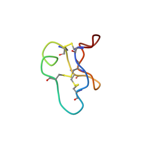
The three-dimensional solution structure of circulin A, a 30 residue polypeptide from the African plant Chassalia parvifolia, has been determined using two-dimensional 1H-NMR spectroscopy. Circulin A was originally identified based upon its inhibition of the cytopathic effects and replication of the human immunodeficiency virus. Structural restraints consisting of 369 interproton distances inferred from nuclear Overhauser effects, and 21 backbone dihedral and nine chi1 angle restraints from spin-spin coupling constants were used as input for simulated annealing calculations and energy minimisation in the program X-PLOR. The final set of 12 structures had mean pairwise rms differences over the whole molecule of 0.91 A for the backbone atom, and 1.68 A for all heavy atoms. For the well-defined region encompassing residues 2-12 and 18-27, the corresponding values were 0.71 and 1.66 A, respectively. Circulin A adopts a compact structure consisting of beta-turns and a distorted segment of triple-stranded beta-sheet. Fluorescence spectroscopy provided additional evidence for a solvent-exposed Trp residue. The molecule is stabilised by three disulfide bonds, two of which form an embedded loop completed by the backbone fragments connecting the cysteine residues. A third disulfide bond threads through the centre of this loop to form a "cystine-knot" motif. This motif is present in a range of other biologically active proteins, including omega-contoxin GVIA and Cucurbita maxima trypsin inhibitor. Circulin A belongs to a novel class of macrocyclic peptides which have been isolated from plants in the Rubiaceae family. The global fold of circulin A is similar to kalata B1, the only member of this class for which a structure has previously been determined.
- Centre for Drug Design and Development, University of Queensland, Brisbane, Queensland, 4072, Australia.
Organizational Affiliation: