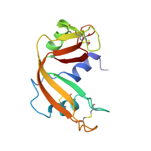
1BEL

HYDROLASE PHOSPHORIC DIESTER, RNA


Experimental Data Snapshot

  • Method: X-RAY DIFFRACTION
  • Resolution: 1.60 Å
  • R-Value Observed: 0.150 

wwPDB Validation   3D Report Full Report


This is version 1.3 of the entry. See complete history


Literature

Structure of crystal form IX of bovine pancreatic ribonuclease A.

Dung, M.H.Bell, J.A.

(1997) Acta Crystallogr D Biol Crystallogr 53: 419-425

  • DOI: https://doi.org/10.1107/S0907444997000929
  • Primary Citation of Related Structures:  
    1BEL

  • PubMed Abstract: 

    The X-ray structure of crystal form IX of bovine pancre- atic ribonuclease A (space group P2(1)2(1)2(1)) is reported at 1.6 A resolution. The structure was refined to an R factor of 15.0% and includes coordinates for two sulfate ions, four methanol molecules and 82 waters. The structure could be superimposed on the highest resolution crystal structure of bovine pancreatic fibonuclease available (in space group P2(1)) with an r.m.s, difference in main-chain atomic positions of 0.51 A. Most of the larger differences between the two structures could be related to crystal lattice contacts. Superposition of the new structure with eight other structures of ribonuclease in six crystal forms resulted in an r.m.s, deviation from the average structure of 0.43 A for all main-chain atoms. This similarity among structures exists in spite of the fact that all nine molecules are in different crystal environments.


  • Organizational Affiliation

    Department of Chemistry and Center for Biophysics, Rensselaer Polytechnic Institute, Troy, NY 12180, USA.


Macromolecules
Find similar proteins by:  (by identity cutoff)  |  3D Structure
Entity ID: 1
MoleculeChains Sequence LengthOrganismDetailsImage
RIBONUCLEASE A124Bos taurusMutation(s): 0 
EC: 3.1.27.5
UniProt
Find proteins for P61823 (Bos taurus)
Explore P61823 
Go to UniProtKB:  P61823
Entity Groups  
Sequence Clusters30% Identity50% Identity70% Identity90% Identity95% Identity100% Identity
UniProt GroupP61823
Sequence Annotations
Expand
  • Reference Sequence
Experimental Data & Validation

Experimental Data

  • Method: X-RAY DIFFRACTION
  • Resolution: 1.60 Å
  • R-Value Observed: 0.150 
  • Space Group: P 21 21 21
Unit Cell:
Length ( Å )Angle ( ˚ )
a = 45.9α = 90
b = 44.8β = 90
c = 52.9γ = 90
Software Package:
Software NamePurpose
bioteXdata collection
bioteXdata reduction
MERLOTphasing
TNTrefinement
bioteXdata scaling

Structure Validation

View Full Validation Report


Entry History 

Deposition Data

Revision History  (Full details and data files)

  • Version 1.0: 1996-10-14
    Type: Initial release
  • Version 1.1: 2008-03-24
    Changes: Version format compliance
  • Version 1.2: 2011-07-13
    Changes: Version format compliance
  • Version 1.3: 2023-08-02
    Changes: Database references, Derived calculations, Refinement description