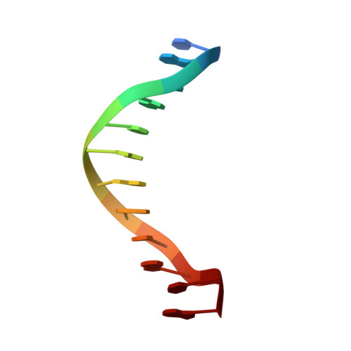
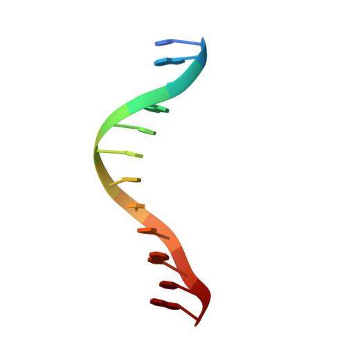
Crystal lattice packing is important in determining the bend of a DNA dodecamer containing an adenine tract.
DiGabriele, A.D., Sanderson, M.R., Steitz, T.A.(1989) Proc Natl Acad Sci U S A 86: 1816-1820
- PubMed: 2928304 Search on PubMedSearch on PubMed Central
- DOI: https://doi.org/10.1073/pnas.86.6.1816
- Primary Citation Related Structures:
1BDN - PubMed Abstract:
The crystal structure of a DNA duplex dodecamer d(CGCAAAAATGCG) and its complementary strand has been determined at 2.6-A resolution. Although our goal was to deduce the structural features of the static bending of the helical axis exhibited by adenine-tract structures in solution, we conclude that the overall bend of 20 degrees in the direction of the major groove observed here arises from the forces associated with crystal packing. An isomorphous dodecamer brominated on one strand provides experimental evidence that this asymmetric sequence is positioned in two orientations in the crystal lattice that are related by a 180 degrees rotation around the pseudodyad axis of the sequence. The bend in these two differently positioned DNA molecules depends on their orientation in the crystal, not on their sequence. As with previously determined structures containing adenine tracts, the adenine and thymine base pairs are highly propeller twisted. The N-6 of the adenine comes within hydrogen bonding distance of the O-4 of thymine one step down the helix, facilitating the formation of a series of bifurcated hydrogen bonds within the adenine tract. The adenine tract is relatively straight and the bending is localized outside this region.
- Department of Chemistry, Yale University, New Haven, CT 06511.
Organizational Affiliation: