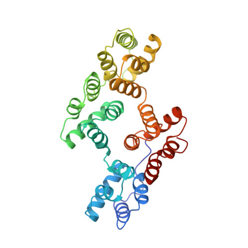
Mutational and crystallographic analyses of interfacial residues in annexin V suggest direct interactions with phospholipid membrane components.
Campos, B., Mo, Y.D., Mealy, T.R., Li, C.W., Swairjo, M.A., Balch, C., Head, J.F., Retzinger, G., Dedman, J.R., Seaton, B.A.(1998) Biochemistry 37: 8004-8010
- PubMed: 9609693 Search on PubMed
- DOI: https://doi.org/10.1021/bi973142n
- Primary Citation Related Structures:
1BC0, 1BC1, 1BC3, 1BCW, 1BCY, 1BCZ - PubMed Abstract:
Annexin V belongs to a family of eukaryotic calcium-dependent membrane-binding proteins. The calcium-binding sites at the annexin-membrane interface have been investigated in some detail; however, little is known about the functional roles of highly conserved interfacial residues that do not coordinate calcium themselves. In the present study, the importance of tryptophan 185, and threonine or serine at positions 72, 144, 228, and 303, in rat annexin V is investigated by site-directed mutagenesis, X-ray crystallography, and functional assays. The high-resolution crystal structures of the mutants show that the mutations do not cause structural perturbations of the annexin molecule itself or disappearance of bound calcium ions from calcium-binding sites. The assays indicate that relative to wild-type annexin V, loss of the methyl substituent at position 72 (Thr72-->Ser) has no effect while loss of the hydroxyl group (Thr72-->Ala or Thr72-->Lys) causes reduction of membrane binding. Multiple lysine substitutions (e.g., Thr72,Ser144,Ser228,Ser303-->Lys) have a greater adverse effect than the single lysine mutation, suggesting that in annexin V the introduction of potentially favorable electrostatic interactions between the lysine side chains and the net negatively charged membrane surface is not sufficient to overcome the loss of the hydroxyl side chains. Replacement of the unique tryptophan, Trp185, by alanine similarly decreases membrane binding affinity. Taken together, the data suggest that the side chains mutated in this study contribute to phospholipid binding and participate directly in intermolecular contacts with phospholipid membrane components.
- Department of Molecular and Cellular Physiology, University of Cincinnati, College of Medicine, Ohio 45220, USA.
Organizational Affiliation: