The use of dipolar couplings for determining the solution structure of rat apo-S100B(betabeta).
Drohat, A.C., Tjandra, N., Baldisseri, D.M., Weber, D.J.(1999) Protein Sci 8: 800-809
- PubMed: 10211826 Search on PubMedSearch on PubMed Central
- DOI: https://doi.org/10.1110/ps.8.4.800
- Primary Citation Related Structures:
1B4C - PubMed Abstract:
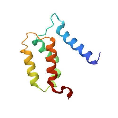
The relative orientations of adjacent structural elements without many well-defined NOE contacts between them are typically poorly defined in NMR structures. For apo-S100B(betabeta) and the structurally homologous protein calcyclin, the solution structures determined by conventional NMR exhibited considerable differences and made it impossible to draw unambiguous conclusions regarding the Ca2+-induced conformational change required for target protein binding. The structure of rat apo-S100B(betabeta) was recalculated using a large number of constraints derived from dipolar couplings that were measured in a dilute liquid crystalline phase. The dipolar couplings orient bond vectors relative to a single-axis system, and thereby remove much of the uncertainty in NOE-based structures. The structure of apo-S100B(betabeta) indicates a minimal change in the first, pseudo-EF-hand Ca2+ binding site, but a large reorientation of helix 3 in the second, classical EF-hand upon Ca2+ binding.
- Department of Biochemistry and Molecular Biology, University of Maryland School of Medicine, Baltimore 21201, USA.
Organizational Affiliation: