1B3U | pdb_00001b3u

CRYSTAL STRUCTURE OF CONSTANT REGULATORY DOMAIN OF HUMAN PP2A, PR65ALPHA


Experimental Data Snapshot

  • Method: X-RAY DIFFRACTION
  • Resolution: 2.30 Å
  • R-Value Free: 
    0.268 (Depositor), 0.265 (DCC) 
  • R-Value Work: 
    0.209 (Depositor), 0.206 (DCC) 

wwPDB Validation 3D Report Full Report

Validation slider image for 1B3U

This is version 1.3 of the entry. See complete history

Literature

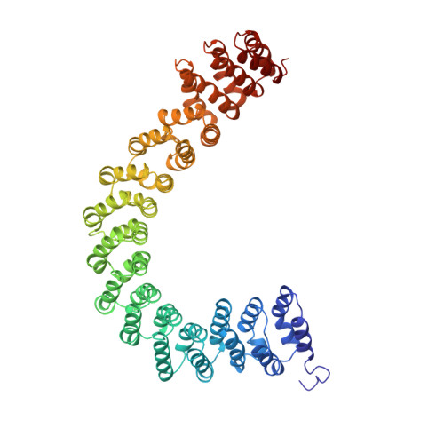
The structure of the protein phosphatase 2A PR65/A subunit reveals the conformation of its 15 tandemly repeated HEAT motifs.

Groves, M.R.Hanlon, N.Turowski, P.Hemmings, B.A.Barford, D.

(1999) Cell 96: 99-110

  • DOI: https://doi.org/10.1016/s0092-8674(00)80963-0
  • Primary Citation Related Structures: 
    1B3U

  • PubMed Abstract: 

    The PR65/A subunit of protein phosphatase 2A serves as a scaffolding molecule to coordinate the assembly of the catalytic subunit and a variable regulatory B subunit, generating functionally diverse heterotrimers. Mutations of the beta isoform of PR65 are associated with lung and colon tumors. The crystal structure of the PR65/Aalpha subunit, at 2.3 A resolution, reveals the conformation of its 15 tandemly repeated HEAT sequences, degenerate motifs of approximately 39 amino acids present in a variety of proteins, including huntingtin and importin beta. Individual motifs are composed of a pair of antiparallel alpha helices that assemble in a mainly linear, repetitive fashion to form an elongated molecule characterized by a double layer of alpha helices. Left-handed rotations at three interrepeat interfaces generate a novel left-hand superhelical conformation. The protein interaction interface is formed from the intrarepeat turns that are aligned to form a continuous ridge.


  • Organizational Affiliation
    • Department of Biochemistry, University of Oxford, United Kingdom.

Macromolecule Content 

  • Total Structure Weight: 130.49 kDa 
  • Atom Count: 9,869 
  • Modeled Residue Count: 1,176 
  • Deposited Residue Count: 1,176 
  • Unique protein chains: 1

Macromolecules

Find similar proteins by:|  3D Structure
Entity ID: 1
MoleculeChains  Sequence LengthOrganismDetailsImage
PROTEIN (PROTEIN PHOSPHATASE PP2A)
A, B
588Homo sapiensMutation(s): 0 
UniProt & NIH Common Fund Data Resources
Find proteins for P30153 (Homo sapiens)
Explore P30153 
Go to UniProtKB:  P30153
PHAROS:  P30153
GTEx:  ENSG00000105568 
Entity Groups
Sequence Clusters30% Identity50% Identity70% Identity90% Identity95% Identity100% Identity
UniProt GroupP30153
Sequence Annotations
Expand
Reference Sequence

Experimental Data & Validation

Experimental Data

  • Method: X-RAY DIFFRACTION
  • Resolution: 2.30 Å
  • R-Value Free:  0.268 (Depositor), 0.265 (DCC) 
  • R-Value Work:  0.209 (Depositor), 0.206 (DCC) 
Space Group: P 1 21 1
Unit Cell:
Length ( Å )Angle ( ˚ )
a = 64.28α = 90
b = 112.15β = 95.6
c = 122.63γ = 90
Software Package:
Software NamePurpose
CNSrefinement
DENZOdata reduction
CCP4data scaling

Structure Validation

View Full Validation Report


Entry History 

Deposition Data

Revision History  (Full details and data files)

  • Version 1.0: 1999-04-12
    Type: Initial release
  • Version 1.1: 2008-04-26
    Changes: Version format compliance
  • Version 1.2: 2011-07-13
    Changes: Version format compliance
  • Version 1.3: 2023-12-27
    Changes: Data collection, Database references