1B3N | pdb_00001b3n

BETA-KETOACYL CARRIER PROTEIN SYNTHASE AS A DRUG TARGET, IMPLICATIONS FROM THE CRYSTAL STRUCTURE OF A COMPLEX WITH THE INHIBITOR CERULENIN.


Experimental Data Snapshot

  • Method: X-RAY DIFFRACTION
  • Resolution: 2.65 Å
  • R-Value Free: 
    0.270 (Depositor), 0.240 (DCC) 
  • R-Value Work: 
    0.213 (Depositor), 0.186 (DCC) 

wwPDB Validation 3D Report Full Report

Validation slider image for 1B3N

Ligand Structure Quality Assessment 


This is version 1.5 of the entry. See complete history

Literature

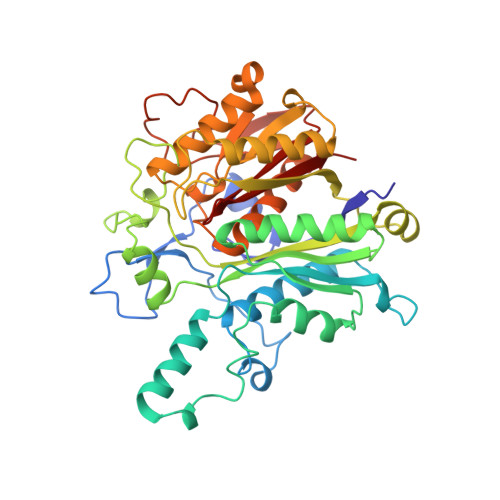
Structure of the complex between the antibiotic cerulenin and its target, beta-ketoacyl-acyl carrier protein synthase.

Moche, M.Schneider, G.Edwards, P.Dehesh, K.Lindqvist, Y.

(1999) J Biological Chem 274: 6031-6034

  • DOI: https://doi.org/10.1074/jbc.274.10.6031
  • Primary Citation Related Structures: 
    1B3N

  • PubMed Abstract: 

    In the biosynthesis of fatty acids, the beta-ketoacyl-acyl carrier protein (ACP) synthases catalyze chain elongation by the addition of two-carbon units derived from malonyl-ACP to an acyl group bound to either ACP or CoA. The enzyme is a possible drug target for treatment of certain cancers and for tuberculosis. The crystal structure of the complex of the enzyme from Escherichia coli, and the fungal mycotoxin cerulenin reveals that the inhibitor is bound in a hydrophobic pocket formed at the dimer interface. Cerulenin is covalently attached to the active site cysteine through its C2 carbon atom. The fit of the inhibitor to the active site is not optimal, and there is thus room for improvement through structure based design.


  • Organizational Affiliation
    • Department of Medical Biochemistry and Biophysics, Doktorsringen 9A1, Karolinska Institutet, S-171 77 Stockholm, Sweden.

Macromolecule Content 

  • Total Structure Weight: 43.18 kDa 
  • Atom Count: 3,074 
  • Modeled Residue Count: 411 
  • Deposited Residue Count: 412 
  • Unique protein chains: 1

Macromolecules

Find similar proteins by:|  3D Structure
Entity ID: 1
MoleculeChains  Sequence LengthOrganismDetailsImage
PROTEIN (KETOACYL ACYL CARRIER PROTEIN SYNTHASE 2)412Escherichia coli K-12Mutation(s): 0 
Gene Names: FABF
EC: 2.3.1.41 (PDB Primary Data), 2.3.1.179 (UniProt)
UniProt
Find proteins for P0AAI5 (Escherichia coli (strain K12))
Explore P0AAI5 
Go to UniProtKB:  P0AAI5
Entity Groups
Sequence Clusters30% Identity50% Identity70% Identity90% Identity95% Identity100% Identity
UniProt GroupP0AAI5
Sequence Annotations
Expand
Reference Sequence

Small Molecules

Ligands 1 Unique
IDChains Name / Formula / InChI Key2D Diagram3D Interactions
CER

Query on CER


Download:Ideal Coordinates CCD File
B [auth A](2S, 3R)-3-HYDROXY-4-OXO-7,10-TRANS,TRANS-DODECADIENAMIDE
C12 H19 N O3
QEPYZBPOTYDXNA-FECJWDPASA-N

Experimental Data & Validation

Experimental Data

  • Method: X-RAY DIFFRACTION
  • Resolution: 2.65 Å
  • R-Value Free:  0.270 (Depositor), 0.240 (DCC) 
  • R-Value Work:  0.213 (Depositor), 0.186 (DCC) 
Space Group: P 31 2 1
Unit Cell:
Length ( Å )Angle ( ˚ )
a = 76.394α = 90
b = 76.394β = 90
c = 147.675γ = 120
Software Package:
Software NamePurpose
SIGMAAmodel building
REFMACrefinement
DENZOdata reduction
CCP4data scaling
SIGMAAphasing

Structure Validation

View Full Validation Report


Ligand Structure Quality Assessment 


Entry History 

Deposition Data

Revision History  (Full details and data files)

  • Version 1.0: 1999-04-06
    Type: Initial release
  • Version 1.1: 2007-10-16
    Changes: Version format compliance
  • Version 1.2: 2011-07-13
    Changes: Derived calculations, Source and taxonomy, Version format compliance
  • Version 1.3: 2018-03-07
    Changes: Data collection
  • Version 1.4: 2023-12-27
    Changes: Data collection, Database references, Derived calculations
  • Version 1.5: 2024-10-16
    Changes: Structure summary