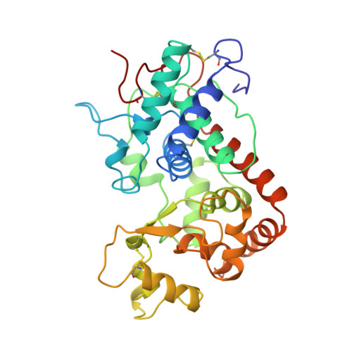
1ATJ

RECOMBINANT HORSERADISH PEROXIDASE C1A


Experimental Data Snapshot

  • Method: X-RAY DIFFRACTION
  • Resolution: 2.15 Å
  • R-Value Free: 0.242 
  • R-Value Work: 0.222 
  • R-Value Observed: 0.222 

Starting Model: experimental
View more details

wwPDB Validation   3D Report Full Report


This is version 1.3 of the entry. See complete history


Literature

Crystal structure of horseradish peroxidase C at 2.15 A resolution.

Gajhede, M.Schuller, D.J.Henriksen, A.Smith, A.T.Poulos, T.L.

(1997) Nat Struct Biol 4: 1032-1038

  • DOI: https://doi.org/10.1038/nsb1297-1032
  • Primary Citation of Related Structures:  
    1ATJ

  • PubMed Abstract: 

    The crystal structure of horseradish peroxidase isozyme C (HRPC) has been solved to 2.15 A resolution. An important feature unique to the class III peroxidases is a long insertion, 34 residues in HRPC, between helices F and G. This region, which defines part of the substrate access channel, is not present in the core conserved fold typical of peroxidases from classes I and II. Comparison of HRPC and peanut peroxidase (PNP), the only other class III (higher plant) peroxidase for which an X-ray structure has been completed, reveals that the structure in this region is highly variable even within class III. For peroxidases of the HRPC type, characterized by a larger FG insertion (seven residues relative to PNP) and a shorter F' helix, we have identified the key residue involved in direct interactions with aromatic donor molecules. HRPC is unique in having a ring of three peripheral Phe residues, 142, 68 and 179. These guard the entrance to the exposed haem edge. We predict that this aromatic region is important for the ability of HRPC to bind aromatic substrates.


  • Organizational Affiliation

    Department of Chemistry, University of Copenhagen, Denmark. gajhede@jerne.ki.ku.dk


Macromolecules
Find similar proteins by:  (by identity cutoff)  |  3D Structure
Entity ID: 1
MoleculeChains Sequence LengthOrganismDetailsImage
PEROXIDASE C1A
A, B, C, D, E
A, B, C, D, E, F
306Armoracia rusticanaMutation(s): 0 
EC: 1.11.1.7
UniProt
Find proteins for P00433 (Armoracia rusticana)
Explore P00433 
Go to UniProtKB:  P00433
Entity Groups  
Sequence Clusters30% Identity50% Identity70% Identity90% Identity95% Identity100% Identity
UniProt GroupP00433
Sequence Annotations
Expand
  • Reference Sequence
Small Molecules
Ligands 2 Unique
IDChains Name / Formula / InChI Key2D Diagram3D Interactions
HEM
Query on HEM

Download Ideal Coordinates CCD File 
I [auth A]
L [auth B]
O [auth C]
R [auth D]
U [auth E]
I [auth A],
L [auth B],
O [auth C],
R [auth D],
U [auth E],
X [auth F]
PROTOPORPHYRIN IX CONTAINING FE
C34 H32 Fe N4 O4
KABFMIBPWCXCRK-RGGAHWMASA-L
CA
Query on CA

Download Ideal Coordinates CCD File 
G [auth A]
H [auth A]
J [auth B]
K [auth B]
M [auth C]
G [auth A],
H [auth A],
J [auth B],
K [auth B],
M [auth C],
N [auth C],
P [auth D],
Q [auth D],
S [auth E],
T [auth E],
V [auth F],
W [auth F]
CALCIUM ION
Ca
BHPQYMZQTOCNFJ-UHFFFAOYSA-N
Experimental Data & Validation

Experimental Data

  • Method: X-RAY DIFFRACTION
  • Resolution: 2.15 Å
  • R-Value Free: 0.242 
  • R-Value Work: 0.222 
  • R-Value Observed: 0.222 
  • Space Group: P 31 1 2
Unit Cell:
Length ( Å )Angle ( ˚ )
a = 159.17α = 90
b = 159.17β = 90
c = 114.27γ = 120
Software Package:
Software NamePurpose
DENZOdata reduction
SCALEPACKdata scaling
X-PLORmodel building
X-PLORrefinement
X-PLORphasing

Structure Validation

View Full Validation Report


Entry History 

Deposition Data

Revision History  (Full details and data files)

  • Version 1.0: 1998-02-04
    Type: Initial release
  • Version 1.1: 2008-03-24
    Changes: Version format compliance
  • Version 1.2: 2011-07-13
    Changes: Version format compliance
  • Version 1.3: 2023-08-02
    Changes: Database references, Derived calculations, Refinement description