1ASX

APICAL DOMAIN OF THE CHAPERONIN FROM THERMOPLASMA ACIDOPHILUM


Experimental Data Snapshot

  • Method: X-RAY DIFFRACTION
  • Resolution: 2.80 Å
  • R-Value Free: 0.272 
  • R-Value Work: 0.222 
  • R-Value Observed: 0.222 

wwPDB Validation   3D Report Full Report


This is version 1.3 of the entry. See complete history


Literature

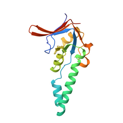
Structure of the substrate binding domain of the thermosome, an archaeal group II chaperonin.

Klumpp, M.Baumeister, W.Essen, L.O.

(1997) Cell 91: 263-270

  • DOI: https://doi.org/10.1016/s0092-8674(00)80408-0
  • Primary Citation of Related Structures:  
    1ASS, 1ASX

  • PubMed Abstract: 

    The crystal structure of the substrate binding domain of the thermosome, the archaeal group II chaperonin, has been determined at 2.3 A resolution. The core resembles the apical domain of GroEL but lacks the hydrophobic residues implied in binding of substrates to group I chaperonins. Rather, a large hydrophobic surface patch is found in a novel helix-turn-helix motif, which is characteristic of all group II chaperonins including the eukaryotic TRiC/CCT complex. Models of the holochaperonin, which are consistent with cryo electron microscopy data, suggest a dual role of this helical protrusion in substrate binding and controlling access to the central cavity independent of a GroES-like cochaperonin.


  • Organizational Affiliation

    Department of Molecular Structural Biology, Max-Planck-Institute for Biochemistry, Planegg-Martinsried, Germany.


Macromolecules
Find similar proteins by:  (by identity cutoff)  |  3D Structure
Entity ID: 1
MoleculeChains Sequence LengthOrganismDetailsImage
THERMOSOME159Thermoplasma acidophilumMutation(s): 0 
Gene Names: THSA
UniProt
Find proteins for P48424 (Thermoplasma acidophilum (strain ATCC 25905 / DSM 1728 / JCM 9062 / NBRC 15155 / AMRC-C165))
Explore P48424 
Go to UniProtKB:  P48424
Entity Groups  
Sequence Clusters30% Identity50% Identity70% Identity90% Identity95% Identity100% Identity
UniProt GroupP48424
Sequence Annotations
Expand
  • Reference Sequence
Small Molecules
Ligands 1 Unique
IDChains Name / Formula / InChI Key2D Diagram3D Interactions
PO4
Query on PO4

Download Ideal Coordinates CCD File 
B [auth A]PHOSPHATE ION
O4 P
NBIIXXVUZAFLBC-UHFFFAOYSA-K
Experimental Data & Validation

Experimental Data

  • Method: X-RAY DIFFRACTION
  • Resolution: 2.80 Å
  • R-Value Free: 0.272 
  • R-Value Work: 0.222 
  • R-Value Observed: 0.222 
  • Space Group: P 31 2 1
Unit Cell:
Length ( Å )Angle ( ˚ )
a = 79.91α = 90
b = 79.91β = 90
c = 70.25γ = 120
Software Package:
Software NamePurpose
SOLOMONphasing
X-PLORmodel building
X-PLORrefinement
DENZOdata reduction
SCALEPACKdata scaling
X-PLORphasing

Structure Validation

View Full Validation Report


Entry History 

Deposition Data

Revision History  (Full details and data files)

  • Version 1.0: 1997-12-03
    Type: Initial release
  • Version 1.1: 2008-03-24
    Changes: Version format compliance
  • Version 1.2: 2011-07-13
    Changes: Derived calculations, Version format compliance
  • Version 1.3: 2023-08-02
    Changes: Database references, Derived calculations, Refinement description