1ARC

THE PRIMARY STRUCTURE AND STRUCTURAL CHARACTERISTICS OF ACHROMOBACTER LYTICUS PROTEASE I, A LYSINE-SPECIFIC SERINE PROTEASE


Experimental Data Snapshot

  • Method: X-RAY DIFFRACTION
  • Resolution: 2.00 Å
  • R-Value Observed: 0.152 

wwPDB Validation   3D Report Full Report


This is version 1.3 of the entry. See complete history


Literature

The primary structure and structural characteristics of Achromobacter lyticus protease I, a lysine-specific serine protease.

Tsunasawa, S.Masaki, T.Hirose, M.Soejima, M.Sakiyama, F.

(1989) J Biol Chem 264: 3832-3839

  • Primary Citation of Related Structures:  
    1ARB, 1ARC

  • PubMed Abstract: 

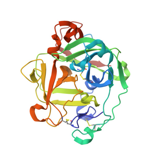
    The complete amino acid sequence of Achromobacter lyticus protease I (EC 3.4.21.50), which specifically hydrolyzes lysyl peptide bonds, has been established. This has been achieved by sequence analysis of the reduced and S-carboxymethylated protease and of peptides obtained by enzymatic digestion with Achromobacter protease I itself and Staphylococcus aureus V8 protease and by chemical cleavage with cyanogen bromide. The protease consists of 268 residues with three disulfide bonds, which have been assigned to Cys6-Cys216, Cys12-Cys80, and Cys36-Cys58. Comparison of the amino acid sequence of Achromobacter protease and other serine proteases of bacterial and mammalian origins has revealed that Achromobacter protease I is a mammalian-type serine protease of which the catalytic triad comprises His57, Asp113, and Ser194. It has also been shown that the protease has 9- and 26-residue extensions of the peptide chain at the N and C termini, respectively, and overall sequence homology is as low as 20% with bovine trypsin. The presence of a disulfide bridge between the N-terminal extension Cys6 and Cys216 close to the putative active site in the C-terminal region is thought to be responsible for the generation of maximal proteolytic function in the pH range 8.5-10.7 and enhanced stability to denaturation.


  • Organizational Affiliation

    Institute for Protein Research, Osaka University, Japan.


Macromolecules
Find similar proteins by:  (by identity cutoff)  |  3D Structure
Entity ID: 1
MoleculeChains Sequence LengthOrganismDetailsImage
ACHROMOBACTER PROTEASE I268Achromobacter lyticusMutation(s): 0 
EC: 3.4.21.50
UniProt
Find proteins for P15636 (Achromobacter lyticus)
Explore P15636 
Go to UniProtKB:  P15636
Entity Groups  
Sequence Clusters30% Identity50% Identity70% Identity90% Identity95% Identity100% Identity
UniProt GroupP15636
Sequence Annotations
Expand
  • Reference Sequence
Small Molecules
Ligands 1 Unique
IDChains Name / Formula / InChI Key2D Diagram3D Interactions
TCK
Query on TCK

Download Ideal Coordinates CCD File 
B [auth A]N-[(1S)-5-amino-1-(chloroacetyl)pentyl]-4-methylbenzenesulfonamide
C14 H21 Cl N2 O3 S
RDFCSSHDJSZMTQ-ZDUSSCGKSA-N
Biologically Interesting Molecules (External Reference) 1 Unique
Entity ID: 2
IDChains NameType/Class2D Diagram3D Interactions
PRD_000459 (TCK)
Query on PRD_000459
B [auth A]Tosyl-L-lysine chloromethyl ketonePeptide-like / Inhibitor
Experimental Data & Validation

Experimental Data

  • Method: X-RAY DIFFRACTION
  • Resolution: 2.00 Å
  • R-Value Observed: 0.152 
  • Space Group: P 1
Unit Cell:
Length ( Å )Angle ( ˚ )
a = 37.3α = 120.1
b = 42.8β = 112.8
c = 48γ = 68.5
Software Package:
Software NamePurpose
PROLSQrefinement

Structure Validation

View Full Validation Report


Entry History 

Deposition Data

Revision History  (Full details and data files)

  • Version 1.0: 1993-10-31
    Type: Initial release
  • Version 1.1: 2008-03-24
    Changes: Version format compliance
  • Version 1.2: 2011-07-13
    Changes: Atomic model, Database references, Derived calculations, Non-polymer description, Structure summary, Version format compliance
  • Version 1.3: 2013-02-27
    Changes: Other