1AOW | pdb_00001aow

ANNEXIN IV


Experimental Data Snapshot

  • Method: X-RAY DIFFRACTION
  • Resolution: 3.00 Å
  • R-Value Free: 
    0.269 (Depositor) 
  • R-Value Work: 
    0.190 (Depositor), 0.206 (DCC) 
  • R-Value Observed: 
    0.190 (Depositor) 

Starting Model: experimental
View more details

wwPDB Validation 3D Report Full Report

Validation slider image for 1AOW

This is version 1.4 of the entry. See complete history

Literature

Structure of the trigonal crystal form of bovine annexin IV.

Zanotti, G.Malpeli, G.Gliubich, F.Folli, C.Stoppini, M.Olivi, L.Savoia, A.Berni, R.

(1998) Biochem J 329: 101-106

  • DOI: https://doi.org/10.1042/bj3290101
  • Primary Citation Related Structures: 
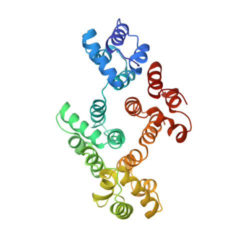
    1AOW

  • PubMed Abstract: 

    The structure of a trigonal crystal form of N-terminally truncated [des-(1-9)] bovine annexin IV, an annexin variant that exhibits the distinctive property of binding both phospholipids and carbohydrates in a Ca2+-dependent manner, has been determined at 3 A (0.3 nm) resolution -space group: R3; cell parameters: a=b=118.560 (8) A and c=82.233 (6) A-. The overall structure of annexin IV, crystallized in the absence of Ca2+ ions, is highly homologous to that of the other known members of the annexin family. The trimeric assembly in the trigonal crystals of annexin IV is quite similar to that found previously in non-isomorphous crystals of human, chicken and rat annexin V and to the subunit arrangement in half of the hexamer of hydra annexin XII. Moreover, it resembles that found in two-dimensional crystals of human annexin V bound to phospholipid monolayers. The propensity of several annexins to generate similar trimeric arrays supports the hypothesis that trimeric complexes of such annexins, including annexin IV, may represent the functional units that interact with membranes.


  • Organizational Affiliation
    • Department of Organic Chemistry, University of Padova, and Biopolymer Research Center, C.N.R., 35131 Padova, Italy.

Macromolecule Content 

  • Total Structure Weight: 35.01 kDa 
  • Atom Count: 2,448 
  • Modeled Residue Count: 309 
  • Deposited Residue Count: 309 
  • Unique protein chains: 1

Macromolecules

Find similar proteins by:|  3D Structure
Entity ID: 1
MoleculeChains  Sequence LengthOrganismDetailsImage
ANNEXIN IV309Bos taurusMutation(s): 0 
Membrane Entity: Yes 
UniProt
Find proteins for P13214 (Bos taurus)
Explore P13214 
Go to UniProtKB:  P13214
Entity Groups
Sequence Clusters30% Identity50% Identity70% Identity90% Identity95% Identity100% Identity
UniProt GroupP13214
Sequence Annotations
Expand
Reference Sequence

Experimental Data & Validation

Experimental Data

  • Method: X-RAY DIFFRACTION
  • Resolution: 3.00 Å
  • R-Value Free:  0.269 (Depositor) 
  • R-Value Work:  0.190 (Depositor), 0.206 (DCC) 
  • R-Value Observed: 0.190 (Depositor) 
Space Group: H 3
Unit Cell:
Length ( Å )Angle ( ˚ )
a = 118.56α = 90
b = 118.56β = 90
c = 82.233γ = 120
Software Package:
Software NamePurpose
X-PLORmodel building
X-PLORrefinement
XDSdata reduction
MARSCALEdata scaling
X-PLORphasing

Structure Validation

View Full Validation Report


Entry History 

Deposition Data

Revision History  (Full details and data files)

  • Version 1.0: 1998-01-14
    Type: Initial release
  • Version 1.1: 2008-03-24
    Changes: Version format compliance
  • Version 1.2: 2011-07-13
    Changes: Version format compliance
  • Version 1.3: 2023-08-02
    Changes: Database references, Other, Refinement description
  • Version 1.4: 2024-05-22
    Changes: Data collection