Structure of a murine leukemia virus receptor-binding glycoprotein at 2.0 angstrom resolution.
Fass, D., Davey, R.A., Hamson, C.A., Kim, P.S., Cunningham, J.M., Berger, J.M.(1997) Science 277: 1662-1666
- PubMed: 9287219 Search on PubMed
- DOI: https://doi.org/10.1126/science.277.5332.1662
- Primary Citation Related Structures:
1AOL - PubMed Abstract:
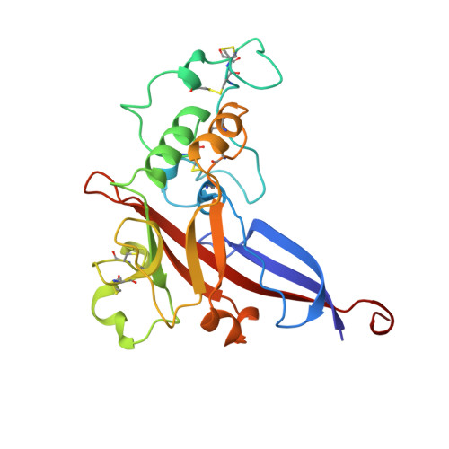
An essential step in retrovirus infection is the binding of the virus to its receptor on a target cell. The structure of the receptor-binding domain of the envelope glycoprotein from Friend murine leukemia virus was determined to 2.0 angstrom resolution by x-ray crystallography. The core of the domain is an antiparallel beta sandwich, with two interstrand loops forming a helical subdomain atop the sandwich. The residues in the helical region, but not in the beta sandwich, are highly variable among mammalian C-type retroviruses with distinct tropisms, indicating that the helical subdomain determines the receptor specificity of the virus.
- Howard Hughes Medical Institute, Whitehead Institute for Biomedical Research, Department of Biology, Massachusetts Institute of Technology, Cambridge, MA 02142, USA.
Organizational Affiliation: