1AOJ | pdb_00001aoj

THE SH3 DOMAIN OF EPS8 EXISTS AS A NOVEL INTERTWINED DIMER


Experimental Data Snapshot

  • Method: X-RAY DIFFRACTION
  • Resolution: 2.50 Å
  • R-Value Free: 
    0.270 (Depositor) 
  • R-Value Work: 
    0.193 (Depositor) 

Starting Model: experimental
View more details

wwPDB Validation 3D Report Full Report

Validation slider image for 1AOJ

This is version 1.4 of the entry. See complete history

Literature

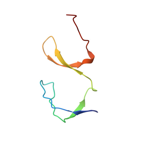
The SH3 domain of Eps8 exists as a novel intertwined dimer.

Kishan, K.V.Scita, G.Wong, W.T.Di Fiore, P.P.Newcomer, M.E.

(1997) Nat Struct Biol 4: 739-743

  • DOI: https://doi.org/10.1038/nsb0997-739
  • Primary Citation Related Structures: 
    1AOJ

  • PubMed Abstract: 

    SH3 domains are structurally well-characterized as monomeric modular units of protein structure that mediate protein-protein recognition in numerous signal transduction proteins. The X-ray crystallographic structure of the Eps8 SH3 domain reveals a novel variation of the canonical SH3 fold: the SH3 domain from Eps8 is a dimer formed by strand interchange. In addition, co-immunoprecipitation experiments show that intact Eps8 is multimeric in vivo. Hence, the SH3 domain of Eps8 may represent a dimerization motif.


  • Organizational Affiliation
    • Department of Biochemistry, Vanderbilt University School of Medicine, Nashville, TN 37232-0146, USA.

Macromolecule Content 

  • Total Structure Weight: 14.09 kDa 
  • Atom Count: 1,019 
  • Modeled Residue Count: 120 
  • Deposited Residue Count: 120 
  • Unique protein chains: 1

Macromolecules

Find similar proteins by:|  3D Structure
Entity ID: 1
MoleculeChains  Sequence LengthOrganismDetailsImage
EPS8
A, B
60Mus musculusMutation(s): 0 
UniProt & NIH Common Fund Data Resources
Find proteins for Q08509 (Mus musculus)
Explore Q08509 
Go to UniProtKB:  Q08509
IMPC:  MGI:104684
Entity Groups
Sequence Clusters30% Identity50% Identity70% Identity90% Identity95% Identity100% Identity
UniProt GroupQ08509
Sequence Annotations
Expand
Reference Sequence

Experimental Data & Validation

Experimental Data

  • Method: X-RAY DIFFRACTION
  • Resolution: 2.50 Å
  • R-Value Free:  0.270 (Depositor) 
  • R-Value Work:  0.193 (Depositor) 
Space Group: P 1
Unit Cell:
Length ( Å )Angle ( ˚ )
a = 27.69α = 107.53
b = 28.338β = 96.83
c = 36.825γ = 104.51
Software Package:
Software NamePurpose
ALMNmodel building
TNTrefinement
DENZOdata reduction
SCALEPACKdata scaling
CCP4phasing

Structure Validation

View Full Validation Report


Entry History 

Deposition Data

Revision History  (Full details and data files)

  • Version 1.0: 1998-07-08
    Type: Initial release
  • Version 1.1: 2008-03-24
    Changes: Version format compliance
  • Version 1.2: 2011-07-13
    Changes: Version format compliance
  • Version 1.3: 2023-08-02
    Changes: Database references, Other, Refinement description
  • Version 1.4: 2024-05-22
    Changes: Data collection