1ANK

THE CLOSED CONFORMATION OF A HIGHLY FLEXIBLE PROTEIN: THE STRUCTURE OF E. COLI ADENYLATE KINASE WITH BOUND AMP AND AMPPNP


Experimental Data Snapshot

  • Method: X-RAY DIFFRACTION
  • Resolution: 2.00 Å
  • R-Value Free: 0.316 
  • R-Value Work: 0.201 
  • R-Value Observed: 0.201 

wwPDB Validation   3D Report Full Report


This is version 1.4 of the entry. See complete history


Literature

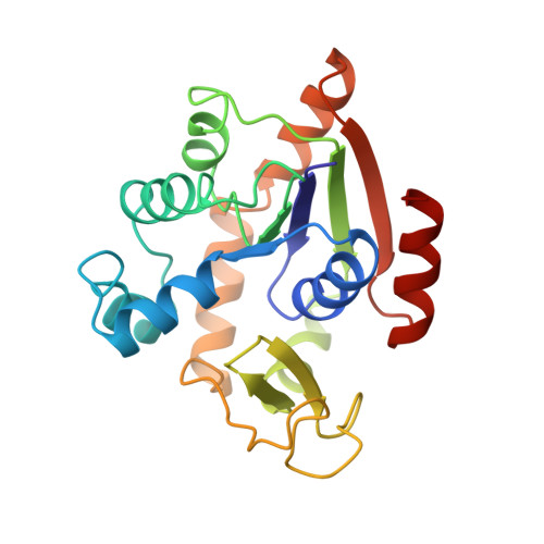
The closed conformation of a highly flexible protein: the structure of E. coli adenylate kinase with bound AMP and AMPPNP.

Berry, M.B.Meador, B.Bilderback, T.Liang, P.Glaser, M.Phillips Jr., G.N.

(1994) Proteins 19: 183-198

  • DOI: https://doi.org/10.1002/prot.340190304
  • Primary Citation of Related Structures:  
    1ANK

  • PubMed Abstract: 

    The structure of E. coli adenylate kinase with bound AMP and AMPPNP at 2.0 A resolution is presented. The protein crystallizes in space group C2 with two molecules in the asymmetric unit, and has been refined to an R factor of 20.1% and an Rfree of 31.6%. In the present structure, the protein is in the closed (globular) form with the large flexible lid domain covering the AMPPNP molecule. Within the protein, AMP and AMPPNP, and ATP analog, occupy the AMP and ATP sites respectively, which had been suggested by the most recent crystal structure of E. coli adenylate kinase with Ap5A bound (Müller and Schulz, 1992, ref. 1) and prior fluorescence studies (Liang et al., 1991, ref. 2). The binding of substrates and the positions of the active site residues are compared between the present structure and the E. coli adenylate kinase/Ap5A structure. We failed to detect a peak in the density map corresponding to the Mg2+ ion which is required for catalysis, and its absence has been attributed to the use of ammonium sulfate in the crystallization solution. Finally, a comparison is made between the present structure and the structure of the heavy chain of muscle myosin.


  • Organizational Affiliation

    W.M. Keck Center for Computational Biology, Rice University, Houston, Texas 77251-1892.


Macromolecules
Find similar proteins by:  (by identity cutoff)  |  3D Structure
Entity ID: 1
MoleculeChains Sequence LengthOrganismDetailsImage
ADENYLATE KINASE
A, B
214Escherichia coliMutation(s): 0 
EC: 2.7.4.3
UniProt
Find proteins for P69441 (Escherichia coli (strain K12))
Explore P69441 
Go to UniProtKB:  P69441
Entity Groups  
Sequence Clusters30% Identity50% Identity70% Identity90% Identity95% Identity100% Identity
UniProt GroupP69441
Sequence Annotations
Expand
  • Reference Sequence
Small Molecules
Ligands 2 Unique
IDChains Name / Formula / InChI Key2D Diagram3D Interactions
ANP
Query on ANP

Download Ideal Coordinates CCD File 
D [auth A],
F [auth B]
PHOSPHOAMINOPHOSPHONIC ACID-ADENYLATE ESTER
C10 H17 N6 O12 P3
PVKSNHVPLWYQGJ-KQYNXXCUSA-N
AMP
Query on AMP

Download Ideal Coordinates CCD File 
C [auth A],
E [auth B]
ADENOSINE MONOPHOSPHATE
C10 H14 N5 O7 P
UDMBCSSLTHHNCD-KQYNXXCUSA-N
Experimental Data & Validation

Experimental Data

  • Method: X-RAY DIFFRACTION
  • Resolution: 2.00 Å
  • R-Value Free: 0.316 
  • R-Value Work: 0.201 
  • R-Value Observed: 0.201 
  • Space Group: C 1 2 1
Unit Cell:
Length ( Å )Angle ( ˚ )
a = 109.7α = 90
b = 73.5β = 131.97
c = 79.6γ = 90
Software Package:
Software NamePurpose
X-PLORmodel building
X-PLORrefinement
X-PLORphasing

Structure Validation

View Full Validation Report


Entry History 

Deposition Data

Revision History  (Full details and data files)

  • Version 1.0: 1994-05-31
    Type: Initial release
  • Version 1.1: 2008-03-24
    Changes: Version format compliance
  • Version 1.2: 2011-07-13
    Changes: Version format compliance
  • Version 1.3: 2017-11-29
    Changes: Derived calculations, Other
  • Version 1.4: 2024-02-07
    Changes: Data collection, Database references, Derived calculations