1AIE | pdb_00001aie

P53 TETRAMERIZATION DOMAIN CRYSTAL STRUCTURE


Experimental Data Snapshot

  • Method: X-RAY DIFFRACTION
  • Resolution: 1.50 Å
  • R-Value Free: 
    0.252 (Depositor), 0.252 (DCC) 
  • R-Value Work: 
    0.191 (Depositor), 0.194 (DCC) 
  • R-Value Observed: 
    0.191 (Depositor) 

Starting Model: experimental
View more details

wwPDB Validation 3D Report Full Report

Validation slider image for 1AIE

This is version 2.1 of the entry. See complete history

Literature

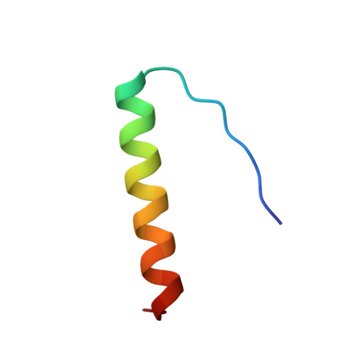
Crystallization and structure solution of p53 (residues 326-356) by molecular replacement using an NMR model as template.

Mittl, P.R.Chene, P.Grutter, M.G.

(1998) Acta Crystallogr D Biol Crystallogr 54: 86-89

  • DOI: https://doi.org/10.1107/s0907444997006550
  • Primary Citation Related Structures: 
    1AIE

  • PubMed Abstract: 

    The molecular replacement method is a powerful technique for crystal structure solution but the use of NMR structures as templates often causes problems. In this work the NMR structure of the p53 tetramerization domain has been used to solve the crystal structure by molecular replacement. Since the rotation- and translation-functions were not sufficiently clear, additional information about the symmetry of the crystal and the protein complex was used to identify correct solutions. The three-dimensional structure of residues 326-356 was subsequently refined to a final R factor of 19.1% at 1.5 A resolution.


  • Organizational Affiliation
    • Core Drug Discovery Technologies, Ciba-Geigy AG, CH-4002 Basel, Switzerland. mittl@fmi.ch

Macromolecule Content 

  • Total Structure Weight: 3.77 kDa 
  • Atom Count: 335 
  • Modeled Residue Count: 31 
  • Deposited Residue Count: 31 
  • Unique protein chains: 1

Macromolecules

Find similar proteins by:|  3D Structure
Entity ID: 1
MoleculeChains  Sequence LengthOrganismDetailsImage
P5331Homo sapiensMutation(s): 0 
UniProt & NIH Common Fund Data Resources
Find proteins for P04637 (Homo sapiens)
Explore P04637 
Go to UniProtKB:  P04637
PHAROS:  P04637
GTEx:  ENSG00000141510 
Entity Groups
Sequence Clusters30% Identity50% Identity70% Identity90% Identity95% Identity100% Identity
UniProt GroupP04637
Sequence Annotations
Expand
Reference Sequence

Experimental Data & Validation

Experimental Data

  • Method: X-RAY DIFFRACTION
  • Resolution: 1.50 Å
  • R-Value Free:  0.252 (Depositor), 0.252 (DCC) 
  • R-Value Work:  0.191 (Depositor), 0.194 (DCC) 
  • R-Value Observed: 0.191 (Depositor) 
Space Group: P 4 2 2
Unit Cell:
Length ( Å )Angle ( ˚ )
a = 45.5α = 90
b = 45.5β = 90
c = 33.2γ = 90
Software Package:
Software NamePurpose
X-PLORmodel building
X-PLORrefinement
XDSdata reduction
MARSCALEdata scaling
X-PLORphasing

Structure Validation

View Full Validation Report


Entry History 

Deposition Data

Revision History  (Full details and data files)

  • Version 1.0: 1997-06-16
    Type: Initial release
  • Version 1.1: 2008-03-24
    Changes: Version format compliance
  • Version 1.2: 2011-07-13
    Changes: Derived calculations, Version format compliance
  • Version 1.3: 2011-11-16
    Changes: Atomic model
  • Version 2.0: 2023-08-02
    Changes: Atomic model, Database references, Other, Refinement description
  • Version 2.1: 2024-05-22
    Changes: Advisory, Data collection, Derived calculations