1A7T | pdb_00001a7t

METALLO-BETA-LACTAMASE WITH MES


Experimental Data Snapshot

  • Method: X-RAY DIFFRACTION
  • Resolution: 1.85 Å
  • R-Value Work: 
    0.151 (Depositor), 0.145 (DCC) 

wwPDB Validation 3D Report Full Report

Validation slider image for 1A7T

This is version 1.4 of the entry. See complete history

Literature

Unanticipated inhibition of the metallo-beta-lactamase from Bacteroides fragilis by 4-morpholineethanesulfonic acid (MES): a crystallographic study at 1.85-A resolution.

Fitzgerald, P.M.Wu, J.K.Toney, J.H.

(1998) Biochemistry 37: 6791-6800

  • DOI: https://doi.org/10.1021/bi9730339
  • Primary Citation Related Structures: 
    1A7T

  • PubMed Abstract: 

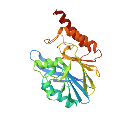
    As part of a structure-aided effort to design clinically useful inhibitors of metallo-beta-lactamases, the X-ray crystal structure of a complex between the metallo-beta-lactamase from Bacteroides fragilis and 4-morpholinoethanesulfonic acid (MES) has been determined and a model for the structure has been refined to a crystallographic R-factor of 0.151 for data between 10.0- and 1.85-A resolution. Although the binding of MES was an adventitious result of the use of MES as a buffer in the crystallization mixture, MES was subsequently shown to be a competitive inhibitor of the enzyme, with a Ki of 23 +/- 5 mM. MES binds in the same fashion to both of the molecules in the crystallographic asymmetric unit; both direct and solvent-mediated hydrogen bonds to the protein and to the binuclear zinc cluster are observed, involving the oxygens of the sulfonic acid group and the nitrogen of the morpholino ring. In addition, there are hydrophobic interactions between the morpholino ring and residues in the flexible beta-strand of the enzyme between residues 26 and 36. Comparison of this structure with the previously reported unliganded structures of the same enzyme [Concha, N. O., Rasmussen, B. A., Bush, K., and Herzberg, O. (1996) Structure 4, 823-836; Carfi, A., Duée, E., Paul-Soto, R., Galleni, M., Frère, J. -M., and Dideberg, O. (1998) Acta Crystallogr. D54, 47-57] reveals that although the overall conservation of structure in the three different crystal lattices is very high, binding of MES is correlated with a significant change in the conformation of this beta-strand. The flexibility of this beta-strand will be an important consideration in the design of inhibitors of the metallo-beta-lactamases.


  • Organizational Affiliation
    • Department of Biochemistry, Merck Research Laboratories, Rahway, New Jersey 07065, USA.

Macromolecule Content 

  • Total Structure Weight: 51.47 kDa 
  • Atom Count: 3,855 
  • Modeled Residue Count: 454 
  • Deposited Residue Count: 464 
  • Unique protein chains: 1

Macromolecules

Find similar proteins by:|  3D Structure
Entity ID: 1
MoleculeChains  Sequence LengthOrganismDetailsImage
METALLO-BETA-LACTAMASE
A, B
232Bacteroides fragilisMutation(s): 2 
Gene Names: CCRA
EC: 3.5.2.6
UniProt
Find proteins for P25910 (Bacteroides fragilis)
Explore P25910 
Go to UniProtKB:  P25910
Entity Groups
Sequence Clusters30% Identity50% Identity70% Identity90% Identity95% Identity100% Identity
UniProt GroupP25910
Sequence Annotations
Expand
Reference Sequence

Small Molecules

Ligands 3 Unique
IDChains Name / Formula / InChI Key2D Diagram3D Interactions
MES

Query on MES


Download:Ideal Coordinates CCD File
F [auth A],
J [auth B]
2-(N-MORPHOLINO)-ETHANESULFONIC ACID
C6 H13 N O4 S
SXGZJKUKBWWHRA-UHFFFAOYSA-N
ZN

Query on ZN


Download:Ideal Coordinates CCD File
C [auth A],
D [auth A],
G [auth B],
H [auth B]
ZINC ION
Zn
PTFCDOFLOPIGGS-UHFFFAOYSA-N
NA

Query on NA


Download:Ideal Coordinates CCD File
E [auth A],
I [auth B]
SODIUM ION
Na
FKNQFGJONOIPTF-UHFFFAOYSA-N

Experimental Data & Validation

Experimental Data

  • Method: X-RAY DIFFRACTION
  • Resolution: 1.85 Å
  • R-Value Work:  0.151 (Depositor), 0.145 (DCC) 
Space Group: P 1 21 1
Unit Cell:
Length ( Å )Angle ( ˚ )
a = 42.27α = 90
b = 66.96β = 103.77
c = 69.48γ = 90
Software Package:
Software NamePurpose
PHASESphasing
PROLSQrefinement
XENGENdata reduction
XENGENdata scaling

Structure Validation

View Full Validation Report


Entry History 

Deposition Data

Revision History  (Full details and data files)

  • Version 1.0: 1998-06-17
    Type: Initial release
  • Version 1.1: 2008-03-24
    Changes: Version format compliance
  • Version 1.2: 2011-07-13
    Changes: Version format compliance
  • Version 1.3: 2021-11-03
    Changes: Database references, Derived calculations
  • Version 1.4: 2024-02-07
    Changes: Data collection