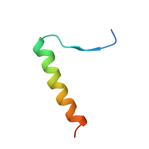
1A1U

SOLUTION STRUCTURE DETERMINATION OF A P53 MUTANT DIMERIZATION DOMAIN, NMR, MINIMIZED AVERAGE STRUCTURE


Experimental Data Snapshot

  • Method: SOLUTION NMR
  • Conformers Calculated: 40 
  • Conformers Submitted: 
  • Selection Criteria: NO VIOLATIONS 

wwPDB Validation   3D Report Full Report

Currently 1A1U does not have a validation slider image.


This is version 1.4 of the entry. See complete history


Literature

Hydrophobic side-chain size is a determinant of the three-dimensional structure of the p53 oligomerization domain.

McCoy, M.Stavridi, E.S.Waterman, J.L.Wieczorek, A.M.Opella, S.J.Halazonetis, T.D.

(1997) EMBO J 16: 6230-6236

  • DOI: https://doi.org/10.1093/emboj/16.20.6230
  • Primary Citation of Related Structures:  
    1A1U

  • PubMed Abstract: 

    The p53 tumor suppressor oligomerization domain, a dimer of two primary dimers, is an independently folding domain whose subunits consist of a beta-strand, a tight turn and an alpha-helix. To evaluate the effect of hydrophobic side-chains on three-dimensional structure, we substituted residues Phe341 and Leu344 in the alpha-helix with other hydrophobic amino acids. Substitutions that resulted in residue 341 having a smaller side-chain than residue 344 switched the stoichiometry of the domain from tetrameric to dimeric. The three-dimensional structure of one such dimer was determined by multidimensional NMR spectroscopy. When compared with the primary dimer of the wild-type p53 oligomerization domain, the mutant dimer showed a switch in alpha-helical packing from anti-parallel to parallel and rotation of the alpha-helices relative to the beta-strands. Hydrophobic side-chain size is therefore an important determinant of a protein fold.


  • Organizational Affiliation

    Departments of Molecular Genetics and Structural Biology, The Wistar Institute, Philadelphia, PA 19104-4268, USA.


Macromolecules
Find similar proteins by:  (by identity cutoff)  |  3D Structure
Entity ID: 1
MoleculeChains Sequence LengthOrganismDetailsImage
P53A,
B [auth C]
35Homo sapiensMutation(s): 0 
Gene Names: P53 TUMOR SUPPRESSOR
UniProt & NIH Common Fund Data Resources
Find proteins for P04637 (Homo sapiens)
Explore P04637 
Go to UniProtKB:  P04637
PHAROS:  P04637
GTEx:  ENSG00000141510 
Entity Groups  
Sequence Clusters30% Identity50% Identity70% Identity90% Identity95% Identity100% Identity
UniProt GroupP04637
Sequence Annotations
Expand
  • Reference Sequence
Experimental Data & Validation

Experimental Data

  • Method: SOLUTION NMR
  • Conformers Calculated: 40 
  • Conformers Submitted: 
  • Selection Criteria: NO VIOLATIONS 

Structure Validation

View Full Validation Report

Currently 1A1U does not have a validation slider image.


Entry History 

Deposition Data

Revision History  (Full details and data files)

  • Version 1.0: 1998-04-08
    Type: Initial release
  • Version 1.1: 2008-03-24
    Changes: Version format compliance
  • Version 1.2: 2011-07-13
    Changes: Version format compliance
  • Version 1.3: 2022-02-16
    Changes: Database references, Derived calculations, Other
  • Version 1.4: 2024-05-22
    Changes: Data collection