1A17 | pdb_00001a17

TETRATRICOPEPTIDE REPEATS OF PROTEIN PHOSPHATASE 5


Experimental Data Snapshot

  • Method: X-RAY DIFFRACTION
  • Resolution: 2.45 Å
  • R-Value Free: 
    0.298 (Depositor) 
  • R-Value Work: 
    0.201 (Depositor), 0.215 (DCC) 
  • R-Value Observed: 
    0.201 (Depositor) 

wwPDB Validation 3D Report Full Report

Validation slider image for 1A17

This is version 1.3 of the entry. See complete history

Literature

The structure of the tetratricopeptide repeats of protein phosphatase 5: implications for TPR-mediated protein-protein interactions.

Das, A.K.Cohen, P.W.Barford, D.

(1998) EMBO J 17: 1192-1199

  • DOI: https://doi.org/10.1093/emboj/17.5.1192
  • Primary Citation Related Structures: 
    1A17

  • PubMed Abstract: 

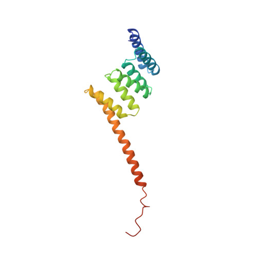
    The tetratricopeptide repeat (TPR) is a degenerate 34 amino acid sequence identified in a wide variety of proteins, present in tandem arrays of 3-16 motifs, which form scaffolds to mediate protein-protein interactions and often the assembly of multiprotein complexes. TPR-containing proteins include the anaphase promoting complex (APC) subunits cdc16, cdc23 and cdc27, the NADPH oxidase subunit p67 phox, hsp90-binding immunophilins, transcription factors, the PKR protein kinase inhibitor, and peroxisomal and mitochondrial import proteins. Here, we report the crystal structure of the TPR domain of a protein phosphatase, PP5. Each of the three TPR motifs of this domain consist of a pair of antiparallel alpha-helices of equivalent length. Adjacent TPR motifs are packed together in a parallel arrangement such that a tandem TPR motif structure is composed of a regular series of antiparallel alpha-helices. The uniform angular and spatial arrangement of neighbouring alpha-helices defines a helical structure and creates an amphipathic groove. Multiple-TPR motif proteins would fold into a right-handed super-helical structure with a continuous helical groove suitable for the recognition of target proteins, hence defining a novel mechanism for protein recognition. The spatial arrangement of alpha-helices in the PP5-TPR domain is similar to those within 14-3-3 proteins.


  • Organizational Affiliation
    • Laboratory of Molecular Biophysics, University of Oxford, Rex Richards Building, South Parks Road, Oxford, OX1 3QU, UK.

Macromolecule Content 

  • Total Structure Weight: 19.25 kDa 
  • Atom Count: 1,369 
  • Modeled Residue Count: 159 
  • Deposited Residue Count: 166 
  • Unique protein chains: 1

Macromolecules

Find similar proteins by:|  3D Structure
Entity ID: 1
MoleculeChains  Sequence LengthOrganismDetailsImage
SERINE/THREONINE PROTEIN PHOSPHATASE 5166Homo sapiensMutation(s): 0 
Gene Names: PPP5
EC: 3.1.3.16
UniProt & NIH Common Fund Data Resources
Find proteins for P53041 (Homo sapiens)
Explore P53041 
Go to UniProtKB:  P53041
PHAROS:  P53041
GTEx:  ENSG00000011485 
Entity Groups
Sequence Clusters30% Identity50% Identity70% Identity90% Identity95% Identity100% Identity
UniProt GroupP53041
Sequence Annotations
Expand
Reference Sequence

Experimental Data & Validation

Experimental Data

  • Method: X-RAY DIFFRACTION
  • Resolution: 2.45 Å
  • R-Value Free:  0.298 (Depositor) 
  • R-Value Work:  0.201 (Depositor), 0.215 (DCC) 
  • R-Value Observed: 0.201 (Depositor) 
Space Group: I 4 2 2
Unit Cell:
Length ( Å )Angle ( ˚ )
a = 90.06α = 90
b = 90.06β = 90
c = 104.45γ = 90
Software Package:
Software NamePurpose
SHARPphasing
X-PLORrefinement
DENZOdata reduction
SCALEPACKdata scaling

Structure Validation

View Full Validation Report


Entry History 

Deposition Data

Revision History  (Full details and data files)

  • Version 1.0: 1998-04-29
    Type: Initial release
  • Version 1.1: 2008-03-24
    Changes: Version format compliance
  • Version 1.2: 2011-07-13
    Changes: Derived calculations, Version format compliance
  • Version 1.3: 2024-02-07
    Changes: Data collection, Database references, Derived calculations