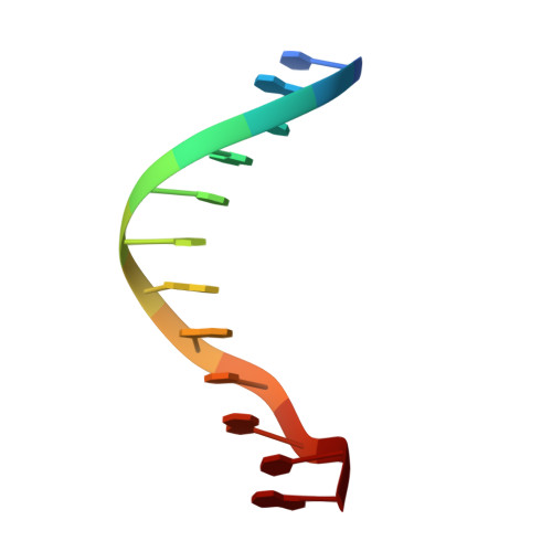
X-ray structures of the B-DNA dodecamer d(CGCGTTAACGCG) with an inverted central tetranucleotide and its netropsin complex.
Balendiran, K., Rao, S.T., Sekharudu, C.Y., Zon, G., Sundaralingam, M.(1995) Acta Crystallogr D Biol Crystallogr 51: 190-198
- PubMed: 15299320
- DOI: https://doi.org/10.1107/S0907444994010759
- Primary Citation of Related Structures:
194D, 195D - PubMed Abstract:
The crystal structures of the B-DNA dodecamer d(CGCGTTAACGCG) duplex (T2A2), with the inverted tetranucleotide core from the duplex d(CGCGAATTCGCG) [A2T2, Dickerson & Drew (1981). J. Mol. Biol. 149, 761-768], and its netropsin complex (T2A2-N) have been determined at 2.3 A resolution. The crystals are orthorhombic, space group P2(1)2(1)2(1), unit-cell dimensions of a = 25.7, b = 40.5 and c = 67.0 A, for T2A2 and a = 25.49, b = 40.87, c = 67.02 A for T2A2-N and are isomorphous with A2T2. The native T2A2 structure, with 70 water molecules had a final R value of 0.15 for 1522 reflections (F > 2sigma), while for the netropsin complex, with 87 water molecules, the R value was 0.16 for 2420 reflections. In T2A2, a discontinuous string of zig-zagging water molecules hydrate the narrow A.T minor groove. In T2A2-N, netropsin binds in one orientation in the minor groove, covering the TTAA central region, by displacing the string of waters, forming the majority of hydrogen bonds with DNA atoms in one strand, and causing very little perturbation of the native structure. The helical twist angle in T2A2 is largest at the duplex center, corresponding to the cleavage site by the restriction enzymes HpaI and HincII. The sequence inversion AATT-->TTAA of the tetranucleotide at the center of the molecule results in a different path for the local helix axis in T2A2 and A2T2 but the overall bending is similar in both cases.
- Department of Biochemistry, College of Agricultural Life Sciences, University of Wisconsin-Madison, 53706, USA.
Organizational Affiliation:


