159D | pdb_0000159d

SIDE BY SIDE BINDING OF TWO DISTAMYCIN A DRUGS IN THE MINOR GROOVE OF AN ALTERNATING B-DNA DUPLEX


Experimental Data Snapshot

  • Method: X-RAY DIFFRACTION
  • Resolution: 1.80 Å
  • R-Value Work: 
    0.150 (Depositor), 0.137 (DCC) 
  • R-Value Observed: 
    0.150 (Depositor) 

wwPDB Validation 3D Report Full Report

Validation slider image for 159D

Ligand Structure Quality Assessment 


This is version 1.3 of the entry. See complete history

Literature

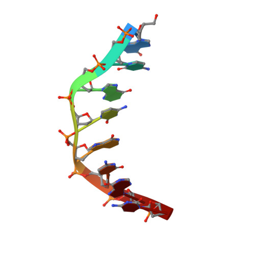
Binding of two distamycin A molecules in the minor groove of an alternating B-DNA duplex.

Chen, X.Ramakrishnan, B.Rao, S.T.Sundaralingam, M.

(1994) Nat Struct Biol 1: 169-175

  • DOI: https://doi.org/10.1038/nsb0394-169
  • Primary Citation Related Structures: 
    159D

  • PubMed Abstract: 

    Here we report the 1.8A X-ray structure of a 2:1 drug-DNA complex between distamycin A and an alternating B-DNA octamer duplex d(ICICICIC)2. The two distamycin A molecules are bound side by side with dyad symmetry in an antiparallel orientation in the expanded minor groove. The amides of each drug molecule are hydrogen bonded to the minor groove base atoms of only one DNA strand. The complex not only shows binding of two drug molecules, but the DNA duplex also exhibits striking low-high alternations in the helical twist angles, the sugar puckering and the phosphate conformations, providing the basis for a new model for an alternating B-DNA with a dinucleotide repeat.


  • Organizational Affiliation
    • Department of Chemistry, Ohio State University, Columbus 43210, USA.

Macromolecule Content 

  • Total Structure Weight: 2.87 kDa 
  • Atom Count: 258 
  • Modeled Residue Count: 8 
  • Deposited Residue Count: 8 
  • Unique nucleic acid chains: 1

Macromolecules

Find similar nucleic acids by:  Sequence
Entity ID: 1
MoleculeChains LengthOrganismImage
DNA (5'-D(*IP*CP*IP*CP*IP*CP*IP*C)-3')8N/A
Sequence Annotations
Expand
Reference Sequence

Experimental Data & Validation

Experimental Data

  • Method: X-RAY DIFFRACTION
  • Resolution: 1.80 Å
  • R-Value Work:  0.150 (Depositor), 0.137 (DCC) 
  • R-Value Observed: 0.150 (Depositor) 
Space Group: P 41 2 2
Unit Cell:
Length ( Å )Angle ( ˚ )
a = 27.93α = 90
b = 27.93β = 90
c = 57.67γ = 90
Software Package:
Software NamePurpose
X-PLORrefinement

Structure Validation

View Full Validation Report


Ligand Structure Quality Assessment 


Entry History 

Deposition Data

Revision History  (Full details and data files)

  • Version 1.0: 1995-02-07
    Type: Initial release
  • Version 1.1: 2008-05-22
    Changes: Version format compliance
  • Version 1.2: 2011-07-13
    Changes: Version format compliance
  • Version 1.3: 2024-02-07
    Changes: Data collection, Database references, Derived calculations