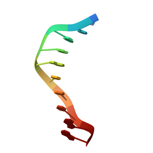
Crystallographic analysis of C-C-A-A-G-C-T-T-G-G and its implications for bending in B-DNA.
Grzeskowiak, K., Goodsell, D.S., Kaczor-Grzeskowiak, M., Cascio, D., Dickerson, R.E.(1993) Biochemistry 32: 8923-8931
- PubMed: 8364037
- DOI: https://doi.org/10.1021/bi00085a025
- Primary Citation of Related Structures:
158D - PubMed Abstract:
Stacked B-DNA double helices of sequence C-C-A-A-G-C-T-T-G-G exhibit the same 23 degrees bend at -T-G-G C-C-A- across the nonbonded junction between helices that is observed in the middle of the decamer helix of sequence C-A-T-G-G-C-C-A-T-G, even though the space group (hexagonal vs orthorhombic), crystal packing, and connectedness at the center of the bent segment are quite different. An identical bend occurs across the interhelix junction of every monoclinic crystal structure of sequence C-C-A-x-x-x-x-T-G-G, suggesting that T-G-G-C-C-A constitutes a natural bending element in B-DNA. The bend occurs by rolling stacked base pairs about their long axes; there is no "tilt" component. Of the three possible models for A-tract bending--bent-A-tract, junction bends, or bent-non-A--which cannot be distinguished by solution measurements, all crystallographic evidence over the past 10 years unanimously supports the non-A regions as the actual bending loci.
- Department of Chemistry and Biochemistry, University of California, Los Angeles 90024.
Organizational Affiliation:


