8AT8

Structure of coproporphyrin III-LmCpfC


Experimental Data Snapshot

  • Method: X-RAY DIFFRACTION
  • Resolution: 1.51 Å
  • R-Value Free: 0.182 
  • R-Value Work: 0.146 
  • R-Value Observed: 0.148 

wwPDB Validation   3D Report Full Report


Ligand Structure Quality Assessment 


This is version 1.2 of the entry. See complete history


Literature

Active site architecture of coproporphyrin ferrochelatase with its physiological substrate coproporphyrin III: Propionate interactions and porphyrin core deformation.

Dali, A.Gabler, T.Sebastiani, F.Destinger, A.Furtmuller, P.G.Pfanzagl, V.Becucci, M.Smulevich, G.Hofbauer, S.

(2023) Protein Sci 32: e4534-e4534

  • DOI: https://doi.org/10.1002/pro.4534
  • Primary Citation of Related Structures:  
    8AT8, 8AW7

  • PubMed Abstract: 

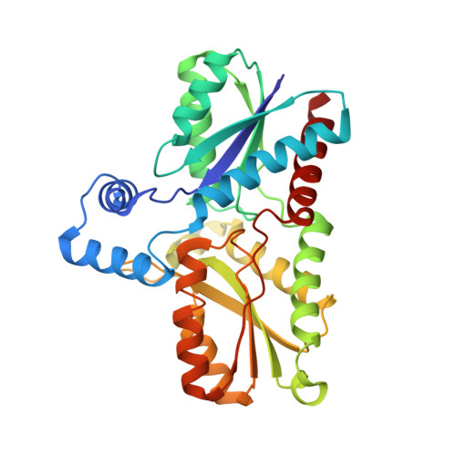
    Coproporphyrin ferrochelatases (CpfCs) are enzymes catalyzing the penultimate step in the coproporphyrin-dependent (CPD) heme biosynthesis pathway, which is mainly utilized by monoderm bacteria. Ferrochelatases insert ferrous iron into a porphyrin macrocycle and have been studied for many decades, nevertheless many mechanistic questions remain unanswered to date. Especially CpfCs, which are found in the CPD pathway, are currently in the spotlight of research. This pathway was identified in 2015 and revealed that the correct substrate for these ferrochelatases is coproporphyrin III (cpIII) instead of protoporphyrin IX, as believed prior the discovery of the CPD pathway. The chemistry of cpIII, which has four propionates, differs significantly from protoporphyrin IX, which features two propionate and two vinyl groups. These findings let us to thoroughly describe the physiological cpIII-ferrochelatase complex in solution and in the crystal phase. Here, we present the first crystallographic structure of the CpfC from the representative monoderm pathogen Listeria monocytogenes bound to its physiological substrate, cpIII, together with the in-solution data obtained by resonance Raman and UV-vis spectroscopy, for wild-type ferrochelatase and variants, analyzing propionate interactions. The results allow us to evaluate the porphyrin distortion and provide an in-depth characterization of the catalytically-relevant binding mode of cpIII prior to iron insertion. Our findings are discussed in the light of the observed structural restraints and necessities for this porphyrin-enzyme complex to catalyze the iron insertion process. Knowledge about this initial situation is essential for understanding the preconditions for iron insertion in CpfCs and builds the basis for future studies.


  • Organizational Affiliation

    Dipartimento di Chimica "Ugo Schiff" - DICUS, Università di Firenze, Sesto Fiorentino (FI), Italy.


Macromolecules
Find similar proteins by:  (by identity cutoff)  |  3D Structure
Entity ID: 1
MoleculeChains Sequence LengthOrganismDetailsImage
Ferrochelatase311Listeria monocytogenesMutation(s): 0 
Gene Names: 
EC: 4.99.1.1
UniProt
Find proteins for Q8Y565 (Listeria monocytogenes serovar 1/2a (strain ATCC BAA-679 / EGD-e))
Explore Q8Y565 
Go to UniProtKB:  Q8Y565
Entity Groups  
Sequence Clusters30% Identity50% Identity70% Identity90% Identity95% Identity100% Identity
UniProt GroupQ8Y565
Sequence Annotations
Expand
  • Reference Sequence
Small Molecules
Ligands 5 Unique
IDChains Name / Formula / InChI Key2D Diagram3D Interactions
HT9 (Subject of Investigation/LOI)
Query on HT9

Download Ideal Coordinates CCD File 
G [auth A]coproporphyrin III
C36 H38 N4 O8
XNBNKCLBGTWWSD-UJJXFSCMSA-N
GOL
Query on GOL

Download Ideal Coordinates CCD File 
E [auth A]GLYCEROL
C3 H8 O3
PEDCQBHIVMGVHV-UHFFFAOYSA-N
EDO
Query on EDO

Download Ideal Coordinates CCD File 
F [auth A]1,2-ETHANEDIOL
C2 H6 O2
LYCAIKOWRPUZTN-UHFFFAOYSA-N
ACT
Query on ACT

Download Ideal Coordinates CCD File 
B [auth A],
C [auth A],
D [auth A]
ACETATE ION
C2 H3 O2
QTBSBXVTEAMEQO-UHFFFAOYSA-M
CA
Query on CA

Download Ideal Coordinates CCD File 
H [auth A]CALCIUM ION
Ca
BHPQYMZQTOCNFJ-UHFFFAOYSA-N
Experimental Data & Validation

Experimental Data

  • Method: X-RAY DIFFRACTION
  • Resolution: 1.51 Å
  • R-Value Free: 0.182 
  • R-Value Work: 0.146 
  • R-Value Observed: 0.148 
  • Space Group: P 1 21 1
Unit Cell:
Length ( Å )Angle ( ˚ )
a = 37.632α = 90
b = 68.069β = 102.224
c = 63.036γ = 90
Software Package:
Software NamePurpose
PHENIXrefinement
PHENIXrefinement
XDSdata reduction
XDSdata scaling
PHASERphasing

Structure Validation

View Full Validation Report


Ligand Structure Quality Assessment 


Entry History & Funding Information

Deposition Data


Funding OrganizationLocationGrant Number
Austrian Science FundAustriaP33544

Revision History  (Full details and data files)

  • Version 1.0: 2022-12-28
    Type: Initial release
  • Version 1.1: 2023-01-11
    Changes: Database references
  • Version 1.2: 2024-01-31
    Changes: Data collection, Refinement description