7YNE

Crystal structure of fragmin domain-1 (1-160) in complex with G-form actin


Experimental Data Snapshot

  • Method: X-RAY DIFFRACTION
  • Resolution: 2.70 Å
  • R-Value Free: 0.308 
  • R-Value Work: 0.273 
  • R-Value Observed: 0.275 

wwPDB Validation   3D Report Full Report


Ligand Structure Quality Assessment 


This is version 1.2 of the entry. See complete history


Literature

Structures and mechanisms of actin ATP hydrolysis.

Kanematsu, Y.Narita, A.Oda, T.Koike, R.Ota, M.Takano, Y.Moritsugu, K.Fujiwara, I.Tanaka, K.Komatsu, H.Nagae, T.Watanabe, N.Iwasa, M.Maeda, Y.Takeda, S.

(2022) Proc Natl Acad Sci U S A 119: e2122641119-e2122641119

  • DOI: https://doi.org/10.1073/pnas.2122641119
  • Primary Citation of Related Structures:  
    7W4Z, 7W50, 7W51, 7W52, 7YNE

  • PubMed Abstract: 

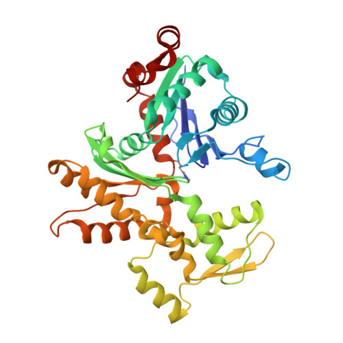
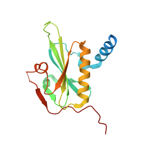
    The major cytoskeleton protein actin undergoes cyclic transitions between the monomeric G-form and the filamentous F-form, which drive organelle transport and cell motility. This mechanical work is driven by the ATPase activity at the catalytic site in the F-form. For deeper understanding of the actin cellular functions, the reaction mechanism must be elucidated. Here, we show that a single actin molecule is trapped in the F-form by fragmin domain-1 binding and present their crystal structures in the ATP analog-, ADP-Pi-, and ADP-bound forms, at 1.15-Å resolutions. The G-to-F conformational transition shifts the side chains of Gln137 and His161, which relocate four water molecules including W1 (attacking water) and W2 (helping water) to facilitate the hydrolysis. By applying quantum mechanics/molecular mechanics calculations to the structures, we have revealed a consistent and comprehensive reaction path of ATP hydrolysis by the F-form actin. The reaction path consists of four steps: 1) W1 and W2 rotations; 2) P G -O 3B bond cleavage; 3) four concomitant events: W1-PO 3 - formation, OH - and proton cleavage, nucleophilic attack by the OH - against P G , and the abstracted proton transfer; and 4) proton relocation that stabilizes the ADP-Pi-bound F-form actin. The mechanism explains the slow rate of ATP hydrolysis by actin and the irreversibility of the hydrolysis reaction. While the catalytic strategy of actin ATP hydrolysis is essentially the same as those of motor proteins like myosin, the process after the hydrolysis is distinct and discussed in terms of Pi release, F-form destabilization, and global conformational changes.


  • Organizational Affiliation

    Graduate School of Information Sciences, Hiroshima City University, Hiroshima 731-3194, Japan.


Macromolecules
Find similar proteins by:  (by identity cutoff)  |  3D Structure
Entity ID: 1
MoleculeChains Sequence LengthOrganismDetailsImage
Actin, alpha skeletal muscle
A, C, E, G
377Gallus gallusMutation(s): 0 
UniProt
Find proteins for P68139 (Gallus gallus)
Explore P68139 
Go to UniProtKB:  P68139
Entity Groups  
Sequence Clusters30% Identity50% Identity70% Identity90% Identity95% Identity100% Identity
UniProt GroupP68139
Sequence Annotations
Expand
  • Reference Sequence
Find similar proteins by:  (by identity cutoff)  |  3D Structure
Entity ID: 2
MoleculeChains Sequence LengthOrganismDetailsImage
Actin-binding protein fragmin P
B, D, F, H
162Physarum polycephalumMutation(s): 0 
UniProt
Find proteins for Q94707 (Physarum polycephalum)
Explore Q94707 
Go to UniProtKB:  Q94707
Entity Groups  
Sequence Clusters30% Identity50% Identity70% Identity90% Identity95% Identity100% Identity
UniProt GroupQ94707
Sequence Annotations
Expand
  • Reference Sequence
Small Molecules
Ligands 6 Unique
IDChains Name / Formula / InChI Key2D Diagram3D Interactions
ADP (Subject of Investigation/LOI)
Query on ADP

Download Ideal Coordinates CCD File 
CA [auth G],
I [auth A],
R [auth C],
Y [auth E]
ADENOSINE-5'-DIPHOSPHATE
C10 H15 N5 O10 P2
XTWYTFMLZFPYCI-KQYNXXCUSA-N
PO4
Query on PO4

Download Ideal Coordinates CCD File 
X [auth D]PHOSPHATE ION
O4 P
NBIIXXVUZAFLBC-UHFFFAOYSA-K
EDO
Query on EDO

Download Ideal Coordinates CCD File 
M [auth A]1,2-ETHANEDIOL
C2 H6 O2
LYCAIKOWRPUZTN-UHFFFAOYSA-N
ACT
Query on ACT

Download Ideal Coordinates CCD File 
K [auth A],
P [auth B],
Q [auth B],
W [auth D]
ACETATE ION
C2 H3 O2
QTBSBXVTEAMEQO-UHFFFAOYSA-M
CA
Query on CA

Download Ideal Coordinates CCD File 
AA [auth F]
BA [auth F]
DA [auth G]
EA [auth H]
FA [auth H]
AA [auth F],
BA [auth F],
DA [auth G],
EA [auth H],
FA [auth H],
J [auth A],
N [auth B],
O [auth B],
S [auth C],
U [auth D],
V [auth D],
Z [auth E]
CALCIUM ION
Ca
BHPQYMZQTOCNFJ-UHFFFAOYSA-N
NA
Query on NA

Download Ideal Coordinates CCD File 
L [auth A],
T [auth C]
SODIUM ION
Na
FKNQFGJONOIPTF-UHFFFAOYSA-N
Modified Residues  1 Unique
IDChains TypeFormula2D DiagramParent
HIC
Query on HIC
A, C, E, G
L-PEPTIDE LINKINGC7 H11 N3 O2HIS
Experimental Data & Validation

Experimental Data

  • Method: X-RAY DIFFRACTION
  • Resolution: 2.70 Å
  • R-Value Free: 0.308 
  • R-Value Work: 0.273 
  • R-Value Observed: 0.275 
  • Space Group: P 21 21 21
Unit Cell:
Length ( Å )Angle ( ˚ )
a = 56.898α = 90
b = 97.999β = 90
c = 420.812γ = 90
Software Package:
Software NamePurpose
PHENIXrefinement
XDSdata reduction
XSCALEdata scaling
MOLREPphasing
PDB_EXTRACTdata extraction

Structure Validation

View Full Validation Report


Ligand Structure Quality Assessment 


Entry History & Funding Information

Deposition Data

  • Released Date: 2022-10-26 
  • Deposition Author(s): Takeda, S.

Funding OrganizationLocationGrant Number
Japan Society for the Promotion of Science (JSPS)Japan16K17708

Revision History  (Full details and data files)

  • Version 1.0: 2022-10-26
    Type: Initial release
  • Version 1.1: 2022-11-02
    Changes: Database references
  • Version 1.2: 2023-11-29
    Changes: Data collection, Refinement description