Serial crystallography captures dynamic control of sequential electron and proton transfer events in a flavoenzyme.
Maestre-Reyna, M., Yang, C.H., Nango, E., Huang, W.C., Ngurah Putu, E.P.G., Wu, W.J., Wang, P.H., Franz-Badur, S., Saft, M., Emmerich, H.J., Wu, H.Y., Lee, C.C., Huang, K.F., Chang, Y.K., Liao, J.H., Weng, J.H., Gad, W., Chang, C.W., Pang, A.H., Sugahara, M., Owada, S., Hosokawa, Y., Joti, Y., Yamashita, A., Tanaka, R., Tanaka, T., Luo, F., Tono, K., Hsu, K.C., Kiontke, S., Schapiro, I., Spadaccini, R., Royant, A., Yamamoto, J., Iwata, S., Essen, L.O., Bessho, Y., Tsai, M.D.(2022) Nat Chem 14: 677-685
- PubMed: 35393554
- DOI: https://doi.org/10.1038/s41557-022-00922-3
- Primary Citation of Related Structures:
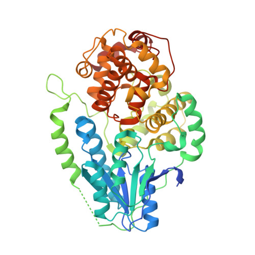
7F8T, 7VIW, 7VIX, 7VIY, 7VIZ, 7VJ0, 7VJ1, 7VJ2, 7VJ3, 7VJ4, 7VJ5, 7VJ6, 7VJ7, 7VJ8, 7VJ9, 7VJA, 7VJB, 7VJC, 7VJE, 7VJG, 7VJH, 7VJI, 7VJJ, 7VJK - PubMed Abstract:
Flavin coenzymes are universally found in biological redox reactions. DNA photolyases, with their flavin chromophore (FAD), utilize blue light for DNA repair and photoreduction. The latter process involves two single-electron transfers to FAD with an intermittent protonation step to prime the enzyme active for DNA repair. Here we use time-resolved serial femtosecond X-ray crystallography to describe how light-driven electron transfers trigger subsequent nanosecond-to-microsecond entanglement between FAD and its Asn/Arg-Asp redox sensor triad. We found that this key feature within the photolyase-cryptochrome family regulates FAD re-hybridization and protonation. After first electron transfer, the FAD •- isoalloxazine ring twists strongly when the arginine closes in to stabilize the negative charge. Subsequent breakage of the arginine-aspartate salt bridge allows proton transfer from arginine to FAD •- . Our molecular videos demonstrate how the protein environment of redox cofactors organizes multiple electron/proton transfer events in an ordered fashion, which could be applicable to other redox systems such as photosynthesis.
Organizational Affiliation:
Institute of Biological Chemistry, Academia Sinica, Nankang, Taipei, Taiwan.